WB
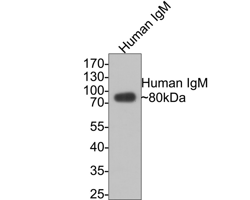
Western blot analysis of Human IgM on human IgM protein with Rabbit anti-Human IgM antibody at 1/500 dilution. Lysates/proteins at 50 ng/Lane. Exposure time: 30 seconds; 10% SDS-PAGE gel. Proteins were transferred to a PVDF membrane and blocked with 5% NFDM/TBST for 1 hour at room temperature. The primary antibody at 1/500 dilution was used in 5% NFDM/TBST at room temperature for 2 hours. Goat Anti-Rabbit IgG - HRP Secondary Antibody at 1:200000 dilution was used for 1 hour at room temperature.ICC/IF
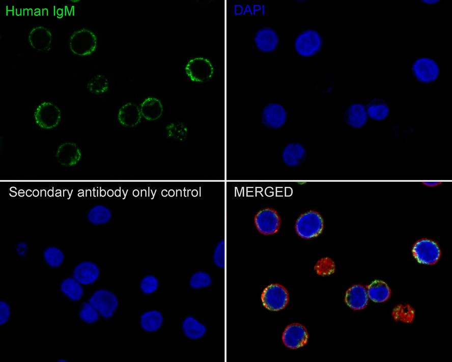
mmunocytochemistry analysis of Daudi cells labeling Human IgM with Rabbit anti-Human IgM antibody at 1/100 dilution. Cells were fixed in 80% precooled methanol for 5 minutes at room temperature, then blocked with 1% BSA in 10% negative goat serum for 1 hour at room temperature. Cells were then incubated with Rabbit anti-Human IgM antibody at 1/100 dilution in 1% BSA in PBST overnight at 4 ℃. Goat Anti-Rabbit IgG H&L (488) was used as the secondary antibody at 1/1000 dilution. PBS instead of the primary antibody was used as the secondary antibody only control. Nuclear DNA was labelled in blue with DAPI. Beta tubulin (red) was stained at 1/100 dilution overnight at +4℃. Goat Anti-Mouse IgG H&L (594) was used as the secondary antibody at 1/1000 dilution.IHC
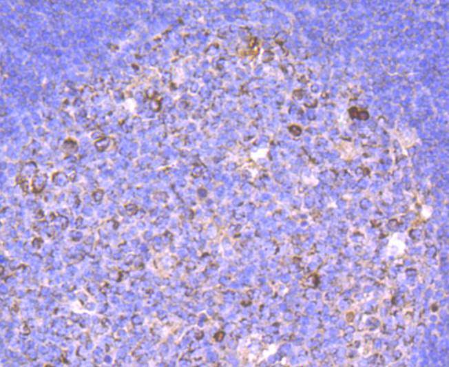
Immunohistochemical analysis of paraffin-embedded human tonsil tissue using anti-Human IgM antibody. The section was pre-treated using heat mediated antigen retrieval with Tris-EDTA buffer (pH 9.0) for 20 minutes.The tissues were blocked in 1% BSA for 30 minutes at room temperature, washed with ddH2O and PBS, and then probed with the primary antibody (1/50) for 30 minutes at room temperature. The detection was performed using an HRP conjugated compact polymer system. DAB was used as the chromogen. Tissues were counterstained with hematoxylin and mounted with DPX.FC
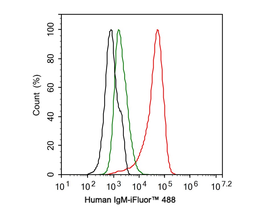
Flow cytometric analysis of Daudi cells labeling Human IgM. Cells were fixed and permeabilized. Then stained with the primary antibody (1μg/mL) (red) compared with Rabbit IgG Isotype Control (green). After incubation of the primary antibody at +4℃ for an hour, the cells were stained with a iFluor™ 488 conjugate-Goat anti-Rabbit IgG Secondary antibody at 1/1000 dilution for 30 minutes at +4℃. Unlabelled sample was used as a control (cells without incubation with primary antibody; black).| Product Name | Human IgM Recombinant Rabbit Monoclonal Antibody |
|---|---|
| Antibody Type | Primary Antibodies |
| Immunogen | Recombinant protein within Human IgM from Human serum (Sigma#I8260). |
| Clonality | monoclonal |
|---|---|
| Isotype | IgG |
| Host Species | Rabbit |
| Tested Applications | FCICC/IFIHCWB |
| WB:1:1000-1:5000 ICC/IF:1:100 IHC:1:50-1:500 FC:1:1000 |
|
| Species Reactivity | Human |
| Concentration | 1mg/ml |
| Purification | Protein A |
| Gene Symbol | IGHM |
|---|---|
| Gene Synonyms | MU VH AGM1 |
| Gene Full Name | immunoglobulin heavy constant mu |
| Gene Summary | Immunoglobulins (Ig) are the antigen recognition molecules of B cells. An Ig molecule is made up of 2 identical heavy chains and 2 identical light chains (see MIM 147200) joined by disulfide bonds so that each heavy chain is linked to a light chain and the 2 heavy chains are linked together. Each Ig heavy chain has an N-terminal variable (V) region containing the antigen-binding site and a C-terminal constant (C) region, encoded by an individual C region gene, that determines the isotype of the antibody and provides effector or signaling functions. The heavy chain V region is encoded by 1 each of 3 types of genes: V genes (see MIM 147070), joining (J) genes (see MIM 147010), and diversity (D) genes (see MIM 146910). The C region genes are clustered downstream of the V region genes within the heavy chain locus on chromosome 14. The IGHM gene encodes the C region of the mu heavy chain, which defines the IgM isotype. Naive B cells express the transmembrane forms of IgM and IgD (see IGHD; MIM 1471770) on their surface. During an antibody response, activated B cells can switch to the expression of individual downstream heavy chain C region genes by a process of somatic recombination known as isotype switching. In addition, secreted Ig forms that act as antibodies can be produced by alternative RNA processing of the heavy chain C region sequences. Although the membrane forms of all Ig isotypes are monomeric, secreted IgM forms pentamers, and occasionally hexamers, in plasma (summary by Janeway et al., 2005).[supplied by OMIM, Aug 2010] |
| Alternative Names | Immunoglobin heavy chain constant region mu edit item name - Immunoglobin heavy chain mu constant region antibody Immunoglobin heavy chain constant region mu antibody AGM1 antibody Constant region of heavy chain of IgM antibody DKFZp686I15196 antibody DKFZp686I15212 antibody FLJ00385 antibody Ig mu chain C region antibody IGHM antibody IgM heavy chain constant region antibody Immunoglobin heavy chain constant region mu edit item name - Immunoglobin heavy chain mu constant region antibody Immunoglobin heavy chain constant region mu antibody AGM1 antibody Constant region of heavy chain of IgM antibody DKFZp686I15196 antibody DKFZp686I15212 antibody FLJ00385 antibody Ig mu chain C region antibody IGHM antibody IgM heavy chain constant region antibody Immunoglobin heavy constant mu antibody Immunoglobulin mu antibody MGC104996 antibody MGC52291 antibody MU antibody VH antibody |
| Molecular Weight(MW) | 50kDa(Observed band size:80kDa) |
| Cellular Localization | Secreted, Cell membrane. |
WB
Western blot analysis of Human IgM on human IgM protein with Rabbit anti-Human IgM antibody at 1/500 dilution. Lysates/proteins at 50 ng/Lane. Exposure time: 30 seconds; 10% SDS-PAGE gel. Proteins were transferred to a PVDF membrane and blocked with 5% NFDM/TBST for 1 hour at room temperature. The primary antibody at 1/500 dilution was used in 5% NFDM/TBST at room temperature for 2 hours. Goat Anti-Rabbit IgG - HRP Secondary Antibody at 1:200000 dilution was used for 1 hour at room temperature.
ICC/IF
mmunocytochemistry analysis of Daudi cells labeling Human IgM with Rabbit anti-Human IgM antibody at 1/100 dilution. Cells were fixed in 80% precooled methanol for 5 minutes at room temperature, then blocked with 1% BSA in 10% negative goat serum for 1 hour at room temperature. Cells were then incubated with Rabbit anti-Human IgM antibody at 1/100 dilution in 1% BSA in PBST overnight at 4 ℃. Goat Anti-Rabbit IgG H&L (488) was used as the secondary antibody at 1/1000 dilution. PBS instead of the primary antibody was used as the secondary antibody only control. Nuclear DNA was labelled in blue with DAPI. Beta tubulin (red) was stained at 1/100 dilution overnight at +4℃. Goat Anti-Mouse IgG H&L (594) was used as the secondary antibody at 1/1000 dilution.
IHC
Immunohistochemical analysis of paraffin-embedded human tonsil tissue using anti-Human IgM antibody. The section was pre-treated using heat mediated antigen retrieval with Tris-EDTA buffer (pH 9.0) for 20 minutes.The tissues were blocked in 1% BSA for 30 minutes at room temperature, washed with ddH2O and PBS, and then probed with the primary antibody (1/50) for 30 minutes at room temperature. The detection was performed using an HRP conjugated compact polymer system. DAB was used as the chromogen. Tissues were counterstained with hematoxylin and mounted with DPX.
FC
Flow cytometric analysis of Daudi cells labeling Human IgM. Cells were fixed and permeabilized. Then stained with the primary antibody (1μg/mL) (red) compared with Rabbit IgG Isotype Control (green). After incubation of the primary antibody at +4℃ for an hour, the cells were stained with a iFluor™ 488 conjugate-Goat anti-Rabbit IgG Secondary antibody at 1/1000 dilution for 30 minutes at +4℃. Unlabelled sample was used as a control (cells without incubation with primary antibody; black).| Application Notes | WB:1:1000-1:5000 ICC/IF:1:100 IHC:1:50-1:500 FC:1:1000 |
|---|
| Form | Liquid |
|---|---|
| Storage Instructions | Shipped at 4℃. Store at +4℃ short term (1-2 weeks). It is recommended to aliquot into single-use upon delivery. Store at -20℃ long term. |
| Storage Buffer | 1*TBS (pH7.4), 0.05% BSA, 40% Glycerol. Preservative: 0.05% Sodium Azide. |


