WB
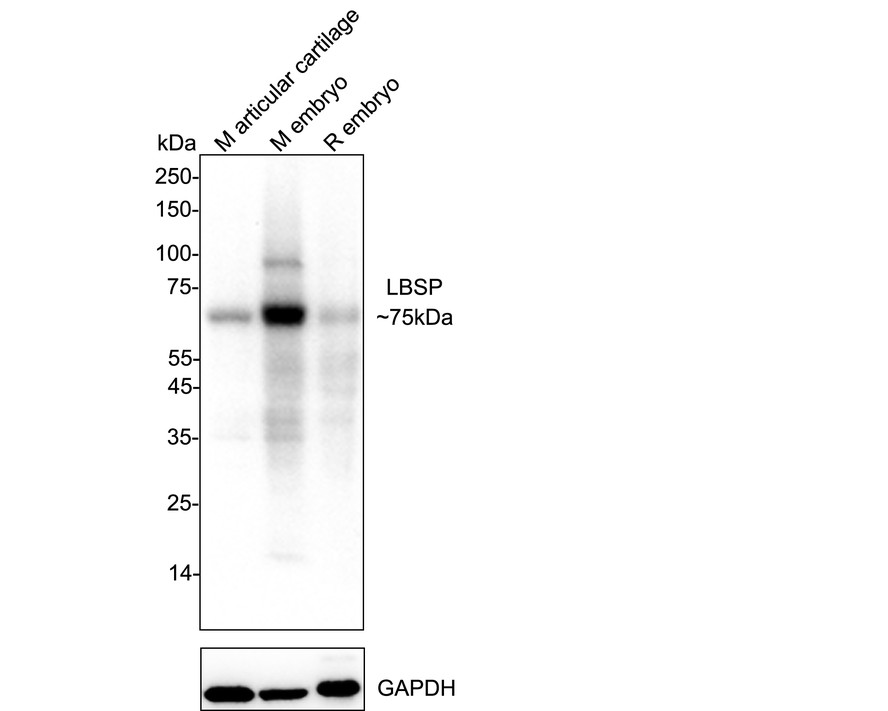
Western blot analysis of Bone sialoprotein 2 on different lysates with Rabbit anti-Bone sialoprotein 2 antibody at 1/2000 dilution. Lane 1: mouse articular cartilage tissue lysate Lane 2: mouse embryo tissue lysate Lane 3: rat embryo tissue lysate Lysates/proteins at 40 µg/Lane. Exposure time: 3 minute ; 4-20% SDS-PAGE gel. Proteins were transferred to a PVDF membrane and blocked with 5% NFDM/TBST for 1 hour at room temperature. The primary antibody at 1/2000 dilution was used in 5% NFDM/TBST at 4℃ overnight. Goat Anti-Rabbit IgG - HRP Secondary Antibody at 1/50000 dilution was used for 1 hour at room temperature.ICC/IF
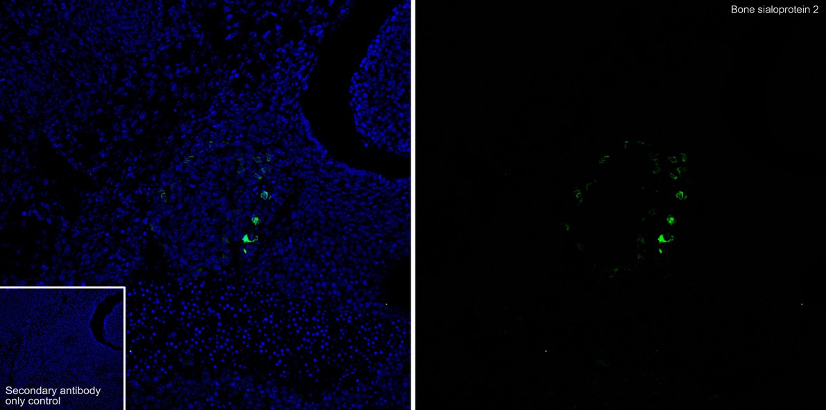
Immunofluorescence analysis of frozen mouse embryonic tissue with Rabbit anti-Bone sialoprotein 2 antibody at 1/500 dilution. The section was not undergone antigen retrieval. The tissues were blocked in 10% negative goat serum for 1 hour at room temperature, washed with PBS, and then probed with the primary antibody (green) at 1/500 dilution overnight at 4 ℃, washed with PBS. Goat Anti-Rabbit IgG H&L (488) was used as the secondary antibody at 1/1000 dilution. Nuclei were counterstained with DAPI (blue).IHC
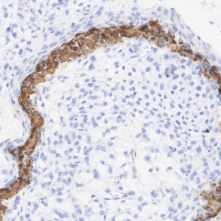
mmunohistochemical analysis of paraffin-embedded mouse embryo tissue with Rabbit anti-Bone sialoprotein 2 antibody at 1/1000 dilution. The section was pre-treated using heat mediated antigen retrieval with Tris-EDTA buffer (pH 9.0) for 20 minutes. The tissues were blocked in 1% BSA for 20 minutes at room temperature, washed with ddH2O and PBS, and then probed with the primary antibody at 1/1000 dilution for 1 hour at room temperature. The detection was performed using an HRP conjugated compact polymer system. DAB was used as the chromogen. Tissues were counterstained with hematoxylin and mounted with DPX.IHC
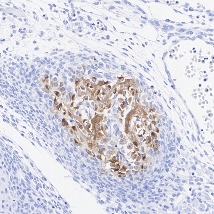
Immunohistochemical analysis of paraffin-embedded rat embryo tissue with Rabbit anti-Bone sialoprotein 2 antibody at 1/1000 dilution. The section was pre-treated using heat mediated antigen retrieval with Tris-EDTA buffer (pH 9.0) for 20 minutes. The tissues were blocked in 1% BSA for 20 minutes at room temperature, washed with ddH2O and PBS, and then probed with the primary antibody at 1/1000 dilution for 1 hour at room temperature. The detection was performed using an HRP conjugated compact polymer system. DAB was used as the chromogen. Tissues were counterstained with hematoxylin and mounted with DPX.| Product Name | Bone sialoprotein 2 Recombinant Rabbit Monoclonal Antibody |
|---|---|
| Antibody Type | Primary Antibodies |
| Product description |
|
| Immunogen | Recombinant protein within mouse Bone sialoprotein 2 aa 1-324/324 |
| Clonality | monoclonal |
|---|---|
| Isotype | IgG |
| Host Species | Rabbit |
| Tested Applications | ICC/IFIHCWB |
| WB:1:2000 ICC/IF:1:500 IHC:1:1000 |
|
| Species Reactivity | MouseRat |
| Concentration | 1mg/ml |
| Purification | Protein A |
| Gene Symbol | Ibsp |
|---|---|
| Gene Synonyms | BSP Bsp2 BSPII BSP II |
| Gene Full Name | integrin binding sialoprotein |
| Gene Summary | The protein encoded by this gene is a major structural protein of the bone matrix. It constitutes approximately 12% of the noncollagenous proteins in human bone and is synthesized by skeletal-associated cell types, including hypertrophic chondrocytes, osteoblasts, osteocytes, and osteoclasts. The only extraskeletal site of its synthesis is the trophoblast. This protein binds to calcium and hydroxyapatite via its acidic amino acid clusters, and mediates cell attachment through an RGD sequence that recognizes the vitronectin receptor. [provided by RefSeq, Jul 2008] |
| Alternative Names | BNSP antibody Bone sialoprotein 2 antibody Bone sialoprotein II antibody BSP antibody BSP II antibody BSPII antibody Cell binding sialoprotein antibody Cell-binding sialoprotein antibody IBSP antibody Integrin binding sialoprotein antibody BNSP antibody Bone sialoprotein 2 antibody Bone sialoprotein II antibody BSP antibody BSP II antibody BSPII antibody Cell binding sialoprotein antibody Cell-binding sialoprotein antibody IBSP antibody Integrin binding sialoprotein antibody Integrin-binding sialoprotein antibody SIAL_HUMAN antibody SPII antibody |
| Molecular Weight(MW) | 70kDa(Observed band size:70kDa) |
| Cellular Localization | Secreted |
WB
Western blot analysis of Bone sialoprotein 2 on different lysates with Rabbit anti-Bone sialoprotein 2 antibody at 1/2000 dilution. Lane 1: mouse articular cartilage tissue lysate Lane 2: mouse embryo tissue lysate Lane 3: rat embryo tissue lysate Lysates/proteins at 40 µg/Lane. Exposure time: 3 minute ; 4-20% SDS-PAGE gel. Proteins were transferred to a PVDF membrane and blocked with 5% NFDM/TBST for 1 hour at room temperature. The primary antibody at 1/2000 dilution was used in 5% NFDM/TBST at 4℃ overnight. Goat Anti-Rabbit IgG - HRP Secondary Antibody at 1/50000 dilution was used for 1 hour at room temperature.
ICC/IF
Immunofluorescence analysis of frozen mouse embryonic tissue with Rabbit anti-Bone sialoprotein 2 antibody at 1/500 dilution. The section was not undergone antigen retrieval. The tissues were blocked in 10% negative goat serum for 1 hour at room temperature, washed with PBS, and then probed with the primary antibody (green) at 1/500 dilution overnight at 4 ℃, washed with PBS. Goat Anti-Rabbit IgG H&L (488) was used as the secondary antibody at 1/1000 dilution. Nuclei were counterstained with DAPI (blue).
IHC
mmunohistochemical analysis of paraffin-embedded mouse embryo tissue with Rabbit anti-Bone sialoprotein 2 antibody at 1/1000 dilution. The section was pre-treated using heat mediated antigen retrieval with Tris-EDTA buffer (pH 9.0) for 20 minutes. The tissues were blocked in 1% BSA for 20 minutes at room temperature, washed with ddH2O and PBS, and then probed with the primary antibody at 1/1000 dilution for 1 hour at room temperature. The detection was performed using an HRP conjugated compact polymer system. DAB was used as the chromogen. Tissues were counterstained with hematoxylin and mounted with DPX.
IHC
Immunohistochemical analysis of paraffin-embedded rat embryo tissue with Rabbit anti-Bone sialoprotein 2 antibody at 1/1000 dilution. The section was pre-treated using heat mediated antigen retrieval with Tris-EDTA buffer (pH 9.0) for 20 minutes. The tissues were blocked in 1% BSA for 20 minutes at room temperature, washed with ddH2O and PBS, and then probed with the primary antibody at 1/1000 dilution for 1 hour at room temperature. The detection was performed using an HRP conjugated compact polymer system. DAB was used as the chromogen. Tissues were counterstained with hematoxylin and mounted with DPX.| Application Notes | WB:1:2000 ICC/IF:1:500 IHC:1:1000 |
|---|
| Form | Liquid |
|---|---|
| Storage Instructions | Shipped at 4℃. Store at +4℃ short term (1-2 weeks). It is recommended to aliquot into single-use upon delivery. Store at -20℃ long term. |
| Storage Buffer | PBS (pH7.4), 0.1% BSA, 40% Glycerol. Preservative: 0.05% Sodium Azide. |


