WB
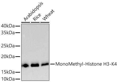
Western blot analysis of various lysates using MonoMethyl-Histone H3-K4 Rabbit mAb at 1:5000dilution incubated overnight at 4℃.Secondary antibody: HRP-conjugated Goat anti-Rabbit IgG (H+L) at 1:10000 dilution.Lysates/proteins: 25 μg per lane.Blocking buffer: 3% nonfat dry milk in TBST.Detection: ECL Basic Kit .Exposure time: 90 s.WB
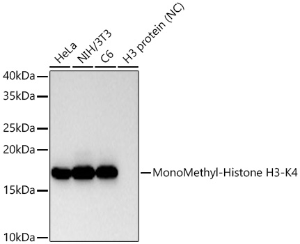
Western blot analysis of various lysates using MonoMethyl-Histone H3-K4 Rabbit mAb at1:10000 dilution incubated overnight at 4℃.Secondary antibody: HRP-conjugated Goat anti-Rabbit IgG (H+L) at 1:10000 dilution.Lysates/proteins: 25 μg per lane.Blocking buffer: 3% nonfat dry milk in TBST.Detection: ECL Basic Kit.Negative control (NC): H3 protein.Exposure time: 45 s.ICC/IF
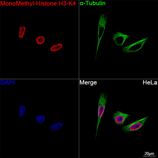
Confocal imaging of HeLa cells usingMonoMethyl-Histone H3-K4 Rabbit mAb(dilution 1:200) followed by afurther incubation with Cy3-conjugated GoatAnti-Rabbit IgG (H+L) (dilution 1:500) (Red). The cells were counterstained with αTubulin Mouse mAb (dilution 1:400)followed by incubation with ABflo® 488-conjugated Goat Anti-Mouse IgG (H+L)(dilution 1:500) (Green). DAPI wasused for nuclear staining (Blue). Objective:100x.ICC/IF
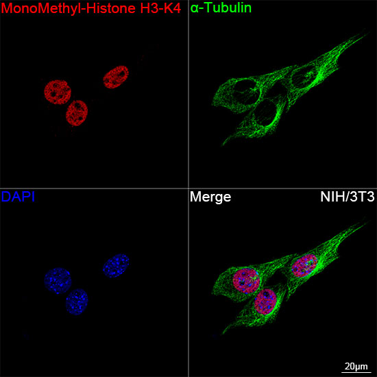
Confocal imaging of NIH/3T3 cells usingMonoMethyl-Histone H3-K4 Rabbit mAb( dilution 1:200) followed by afurther incubation with Cy3-conjugated GoatAnti-Rabbit IgG (H+L) (dilution 1:500) (Red). The cells were counterstained with αTubulin Mouse mAb (dilution 1:400)followed by incubation with ABflo® 488-conjugated Goat Anti-Mouse IgG (H+L)(dilution 1:500) (Green). DAPI wasused for nuclear staining (Blue). Objective:100x.ICC/IF
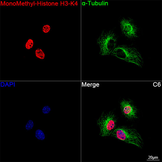
Confocal imaging of C6 cells usingMonoMethyl-Histone H3-K4 Rabbit mAb(dilution 1:200) followed by afurther incubation with Cy3-conjugated GoatAnti-Rabbit IgG (H+L) (dilution 1:500)(Red). The cells were counterstained with αTubulin Mouse mAb (dilution 1:400)followed by incubation with ABflo® 488-conjugated Goat Anti-Mouse IgG (H+L)(dilution 1:500) (Green). DAPI wasused for nuclear staining (Blue). Objective:100x.IHC
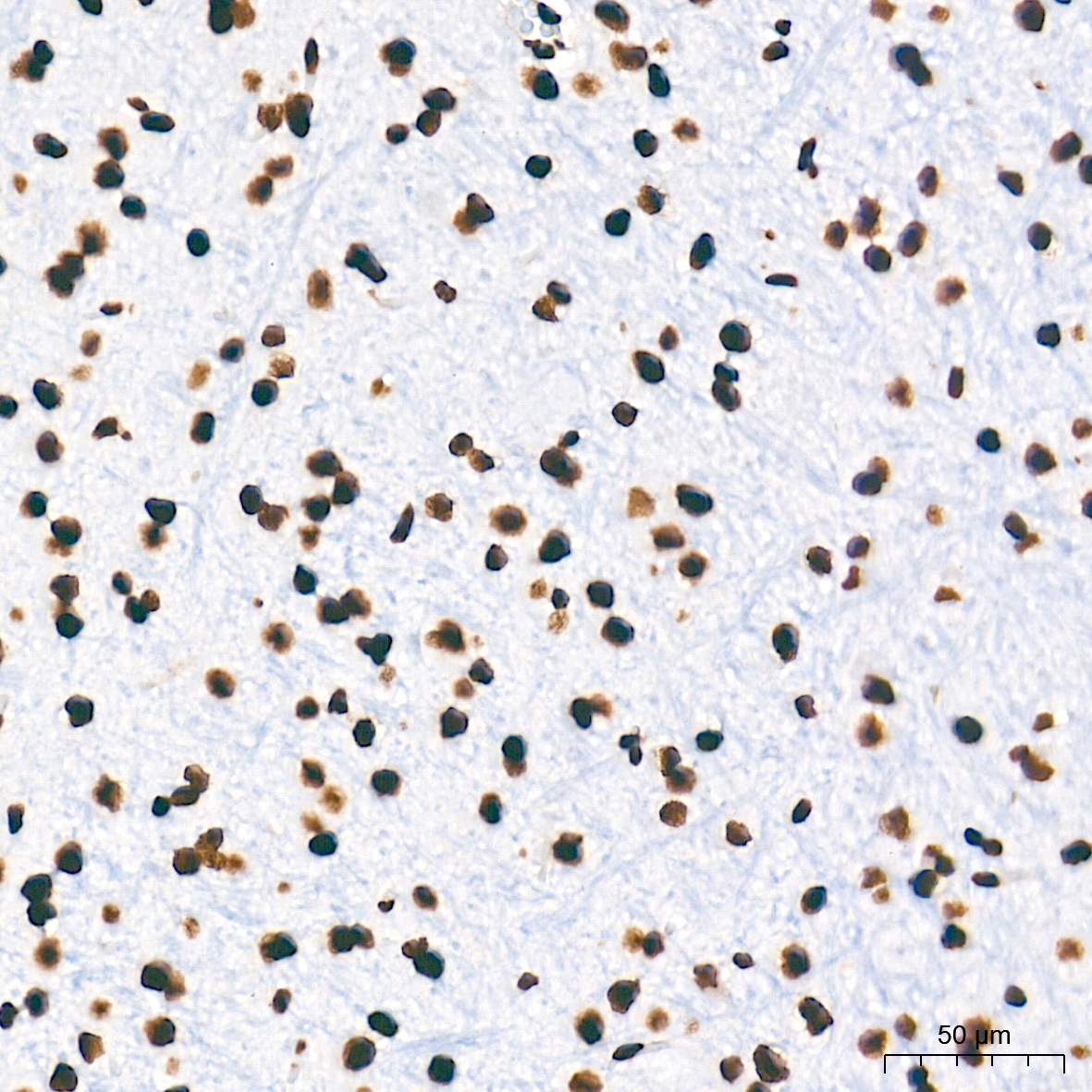
Immunohistochemistry analysis of paraffinembedded Mouse brain tissue usingMonoMethyl-Histone H3-K4 Rabbit mAb at a dilution of 1:16000 (40x lens).High pressure antigen retrieval performedwith 0.01M Tris-EDTA Buffer (pH 9.0) prior toIHC staining.IHC
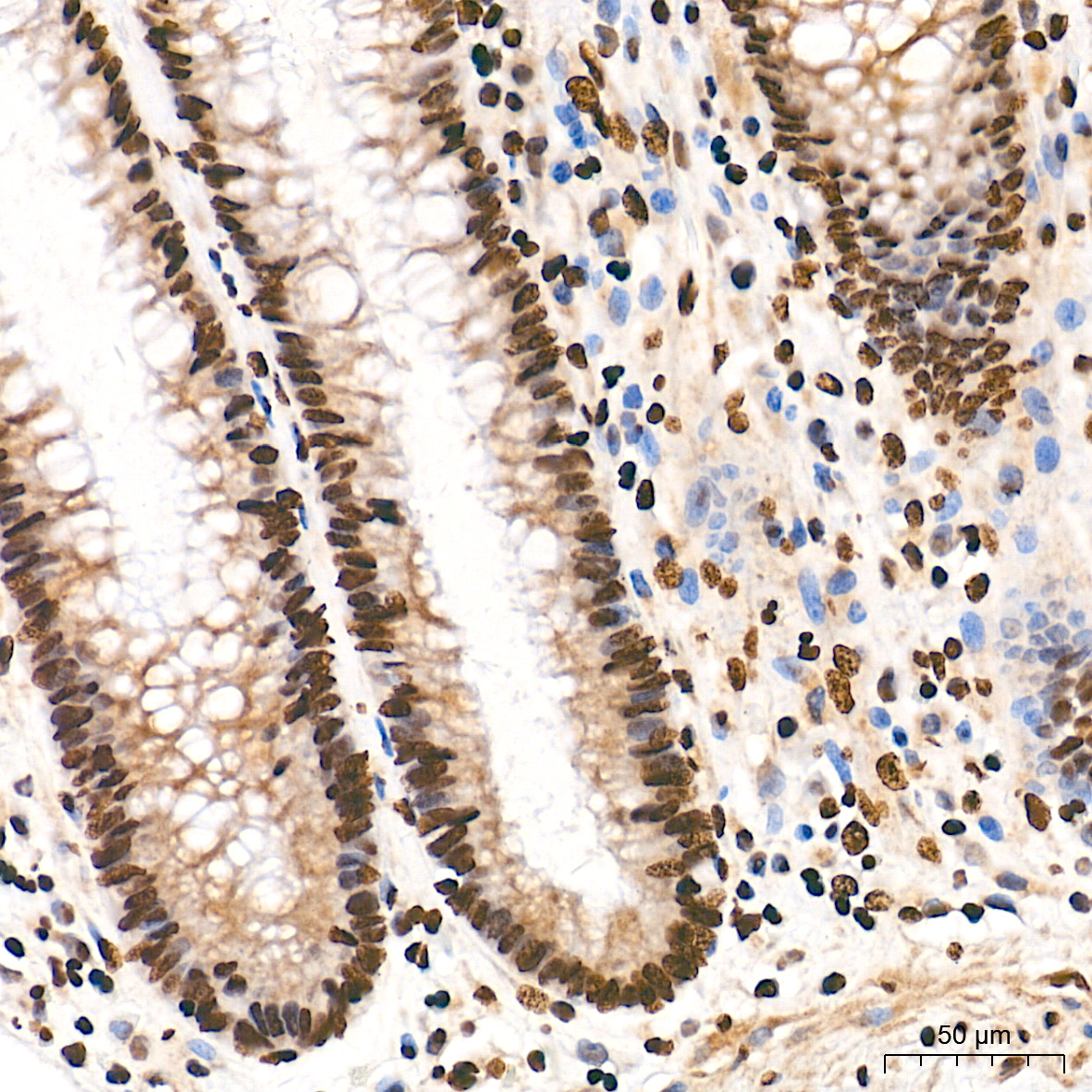
Immunohistochemistry analysis of paraffinembedded Human colon tissue usingMonoMethyl-Histone H3-K4 Rabbit mAb at a dilution of 1:16000 (40x lens).High pressure antigen retrieval performedwith 0.01M Tris-EDTA Buffer (pH 9.0) prior toIHC staining.IHC
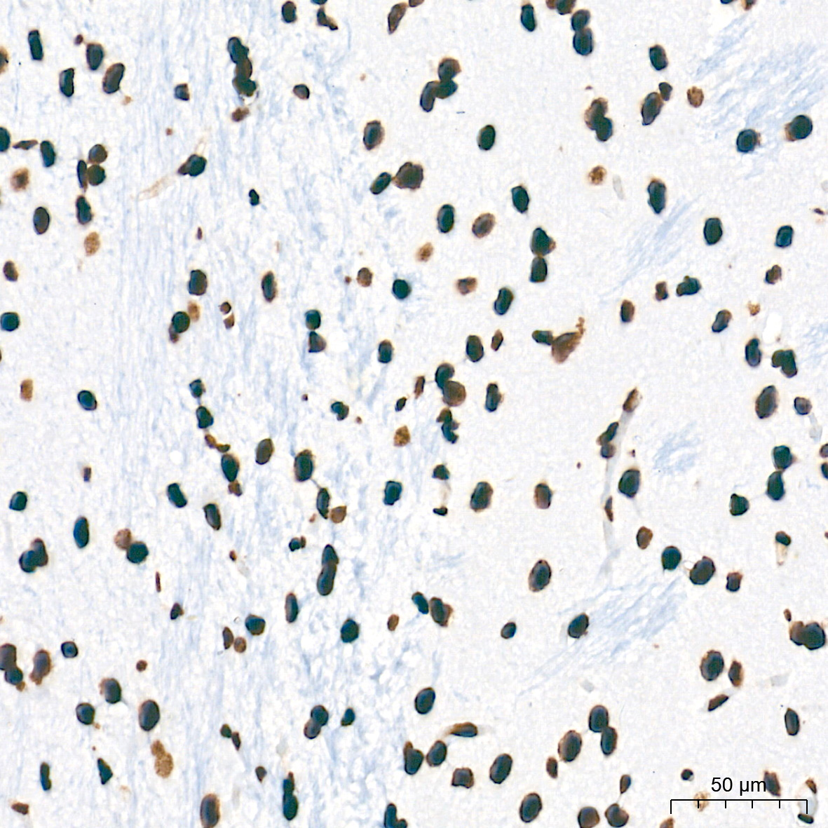
Immunohistochemistry analysis of paraffinembedded Rat brain tissue usingMonoMethyl-Histone H3-K4 Rabbit mAb at a dilution of 1:16000 (40x lens).High pressure antigen retrieval performedwith 0.01M Tris-EDTA Buffer (pH 9.0) prior toIHC staining.ChIP
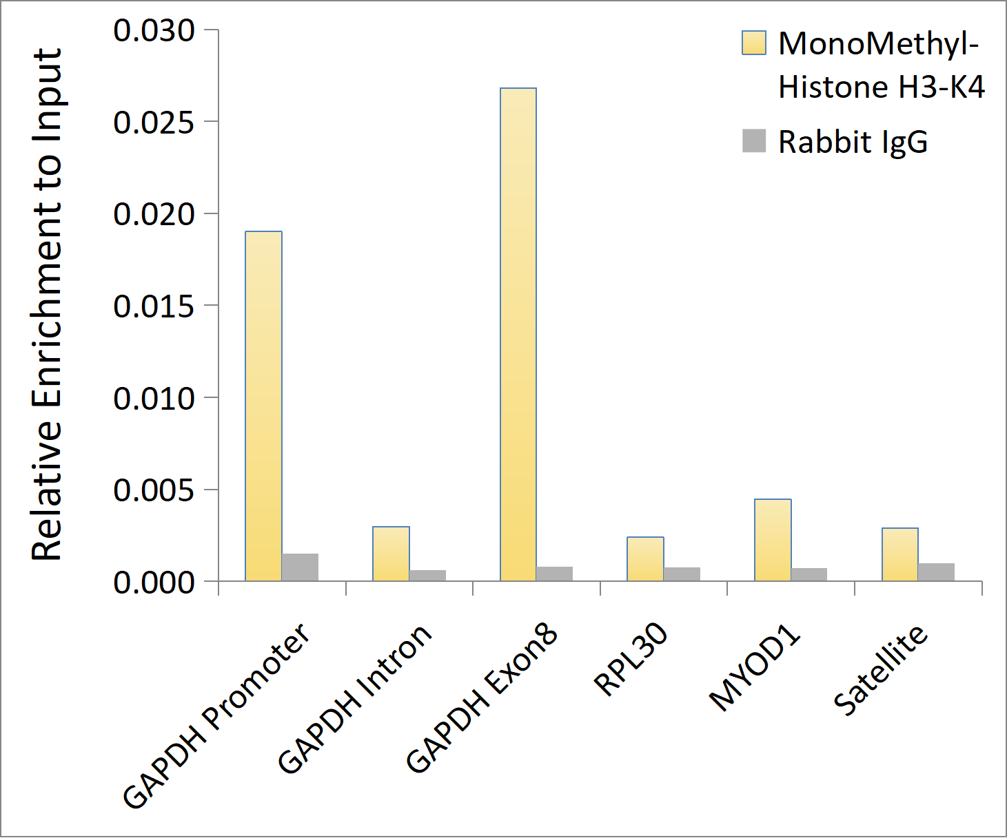
Chromatin immunoprecipitation was performed with 10 μg of cross-linked chromatin from HeLa, using 0.5μg of MonoMethyl-Histone H3-K4 Rabbit mAb and Rabbit IgG isotype control. Theenrichment of immunoprecipitated DNA at different genomic loci was examined by quantitative PCR. Thehistogram compares the ratio of the immunoprecipitated DNA to the input at given loci.DB
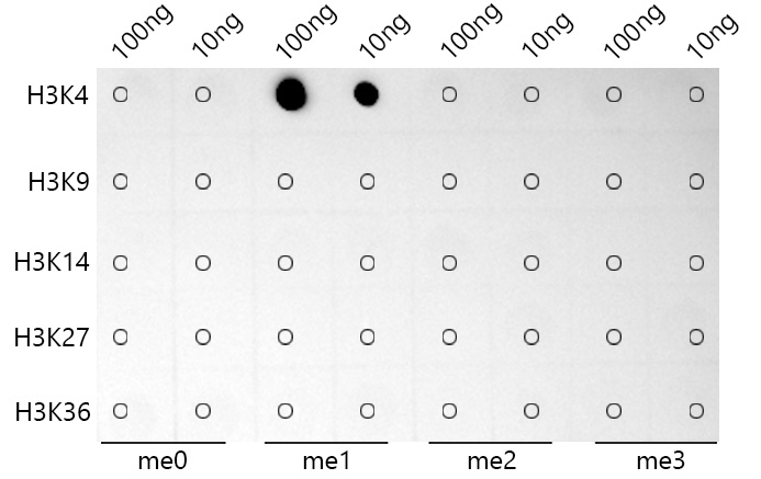
Dot-blot analysis of all sorts of peptidesusing MonoMethyl-Histone H3-K4 Rabbit mAb at 1:1000 dilution.| Product Name | MonoMethyl-Histone H3-K4 Rabbit mAb |
|---|---|
| Antibody Type | Primary Antibodies |
| Immunogen | Synthetic peptide. This information is considered to be commercially sensitive. |
| Modification | MonoMethyl-K4 |
| Clonality | Monoclonal |
|---|---|
| Isotype | IgG |
| Host Species | Rabbit |
| Tested Applications | ChIPDBICC/IFIHCWB |
| WB:1:4000-1:20000 ICC/IF:1:200-1:1000 IHC:1:5000- 1:20000 ChIP:0.5μg antibody for 10μg-15μg of Chromatin DB:1:1000-1:2000 |
|
| Species Reactivity | HumanMouseOtherRatRice |
| Concentration | 1mg/ml |
| Purification | Affinity purified |
| Gene Symbol | H3-4 |
|---|---|
| Gene Synonyms | H3t H3.4 H3/g H3FT H3C16 HIST3H3 |
| Gene Full Name | H3.4 histone, cluster member |
| Gene Summary | Histones are basic nuclear proteins that are responsible for the nucleosome structure of the chromosomal fiber in eukaryotes. Nucleosomes consist of approximately 146 bp of DNA wrapped around a histone octamer composed of pairs of each of the four core histones (H2A, H2B, H3, and H4). The chromatin fiber is further compacted through the interaction of a linker histone, H1, with the DNA between the nucleosomes to form higher order chromatin structures. This gene is intronless and encodes a replication-dependent histone that is a member of the histone H3 family. Transcripts from this gene lack polyA tails; instead, they contain a palindromic termination element. This gene is located separately from the other H3 genes that are in the histone gene cluster on chromosome 6p22-p21.3. [provided by RefSeq, Aug 2015] |
| Alternative Names | H3/A H3C2 H3C3 H3C4 H3C6 H3C7 H3C8 H3FA H3C10 H3C11 H3C12 HIST1H3A Histone H3 H3-4 H3/t H3/g H3FT HIST3H3 |
| Molecular Weight(MW) | 16kDa(Observed band size:17kDa) |
| Cellular Localization | Chromosome, nucleus. |
WB
Western blot analysis of various lysates using MonoMethyl-Histone H3-K4 Rabbit mAb at 1:5000dilution incubated overnight at 4℃.Secondary antibody: HRP-conjugated Goat anti-Rabbit IgG (H+L) at 1:10000 dilution.Lysates/proteins: 25 μg per lane.Blocking buffer: 3% nonfat dry milk in TBST.Detection: ECL Basic Kit .Exposure time: 90 s.
WB
Western blot analysis of various lysates using MonoMethyl-Histone H3-K4 Rabbit mAb at1:10000 dilution incubated overnight at 4℃.Secondary antibody: HRP-conjugated Goat anti-Rabbit IgG (H+L) at 1:10000 dilution.Lysates/proteins: 25 μg per lane.Blocking buffer: 3% nonfat dry milk in TBST.Detection: ECL Basic Kit.Negative control (NC): H3 protein.Exposure time: 45 s.
ICC/IF
Confocal imaging of HeLa cells usingMonoMethyl-Histone H3-K4 Rabbit mAb(dilution 1:200) followed by afurther incubation with Cy3-conjugated GoatAnti-Rabbit IgG (H+L) (dilution 1:500) (Red). The cells were counterstained with αTubulin Mouse mAb (dilution 1:400)followed by incubation with ABflo® 488-conjugated Goat Anti-Mouse IgG (H+L)(dilution 1:500) (Green). DAPI wasused for nuclear staining (Blue). Objective:100x.
ICC/IF
Confocal imaging of NIH/3T3 cells usingMonoMethyl-Histone H3-K4 Rabbit mAb( dilution 1:200) followed by afurther incubation with Cy3-conjugated GoatAnti-Rabbit IgG (H+L) (dilution 1:500) (Red). The cells were counterstained with αTubulin Mouse mAb (dilution 1:400)followed by incubation with ABflo® 488-conjugated Goat Anti-Mouse IgG (H+L)(dilution 1:500) (Green). DAPI wasused for nuclear staining (Blue). Objective:100x.
ICC/IF
Confocal imaging of C6 cells usingMonoMethyl-Histone H3-K4 Rabbit mAb(dilution 1:200) followed by afurther incubation with Cy3-conjugated GoatAnti-Rabbit IgG (H+L) (dilution 1:500)(Red). The cells were counterstained with αTubulin Mouse mAb (dilution 1:400)followed by incubation with ABflo® 488-conjugated Goat Anti-Mouse IgG (H+L)(dilution 1:500) (Green). DAPI wasused for nuclear staining (Blue). Objective:100x.
IHC
Immunohistochemistry analysis of paraffinembedded Mouse brain tissue usingMonoMethyl-Histone H3-K4 Rabbit mAb at a dilution of 1:16000 (40x lens).High pressure antigen retrieval performedwith 0.01M Tris-EDTA Buffer (pH 9.0) prior toIHC staining.
IHC
Immunohistochemistry analysis of paraffinembedded Human colon tissue usingMonoMethyl-Histone H3-K4 Rabbit mAb at a dilution of 1:16000 (40x lens).High pressure antigen retrieval performedwith 0.01M Tris-EDTA Buffer (pH 9.0) prior toIHC staining.
IHC
Immunohistochemistry analysis of paraffinembedded Rat brain tissue usingMonoMethyl-Histone H3-K4 Rabbit mAb at a dilution of 1:16000 (40x lens).High pressure antigen retrieval performedwith 0.01M Tris-EDTA Buffer (pH 9.0) prior toIHC staining.
ChIP
Chromatin immunoprecipitation was performed with 10 μg of cross-linked chromatin from HeLa, using 0.5μg of MonoMethyl-Histone H3-K4 Rabbit mAb and Rabbit IgG isotype control. Theenrichment of immunoprecipitated DNA at different genomic loci was examined by quantitative PCR. Thehistogram compares the ratio of the immunoprecipitated DNA to the input at given loci.
DB
Dot-blot analysis of all sorts of peptidesusing MonoMethyl-Histone H3-K4 Rabbit mAb at 1:1000 dilution.| Application Notes | WB:1:4000-1:20000 ICC/IF:1:200-1:1000 IHC:1:5000- 1:20000 ChIP:0.5μg antibody for 10μg-15μg of Chromatin DB:1:1000-1:2000 |
|---|
| Form | Liquid |
|---|---|
| Storage Instructions | Store at -20℃. Avoid freeze / thaw cycles. |
| Storage Buffer | PBS with 0.09% Sodium azide, 0.05% BSA, 50% glycerol, pH7.3. |


