WB
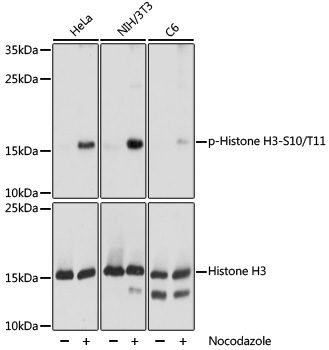
Western blot analysis of lysates from NIH/3T3 cells, using Phospho-Histone H3-S10/T11 Rabbit pAb at 1:1000 dilution. HeLa,NIH/3T3 and C6 cellls were treated with nocodazole (50 ng/mL) at 37℃for 20 hours.Secondary antibody: HRP-conjugated Goat anti-Rabbit IgG (H+L) at 1:10000 dilution.Lysates/proteins: 25μg per lane.Blocking buffer: 3% BSA.Detection: ECL Basic Kit.Exposure time: 1s.WB
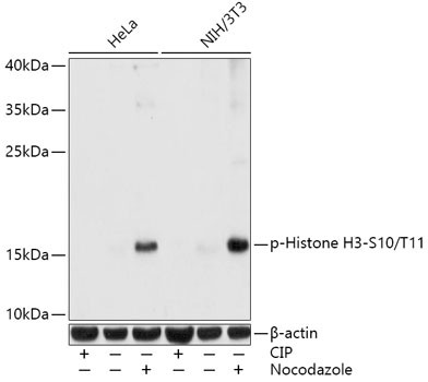
Western blot analysis of various lysates using Phospho-Histone H3-S10/T11 Rabbit pAb at1:1000 dilution. Both NIH/3T3 cells and Hela cells were treated with CIP(20uL/400ul) at 37℃ for 1 hourand treated with nocodazole (50 ng/mL) at 37℃ for 20 hours.Secondary antibody: HRP-conjugated Goat anti-Rabbit IgG (H+L) at 1:10000 dilution.Lysates/proteins: 25μg per lane.Blocking buffer: 3% BSA.Detection: ECL Basic Kit.Exposure time: 1s.ICC/IF
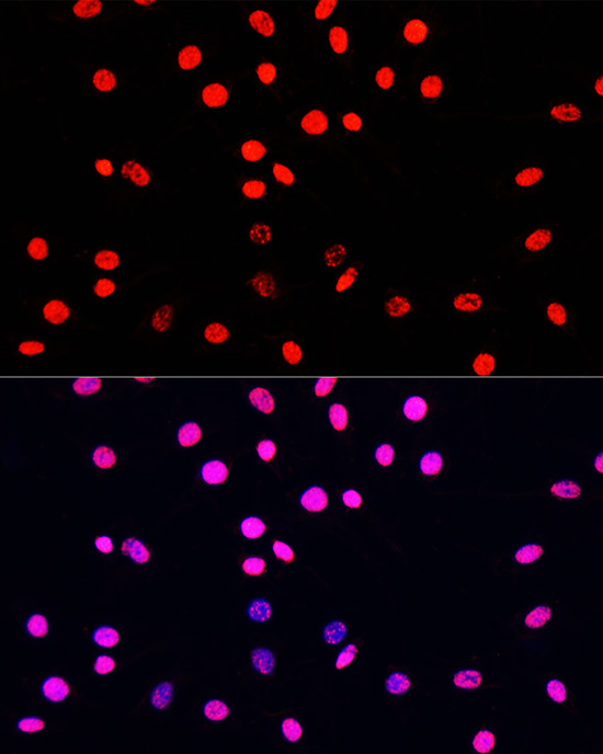
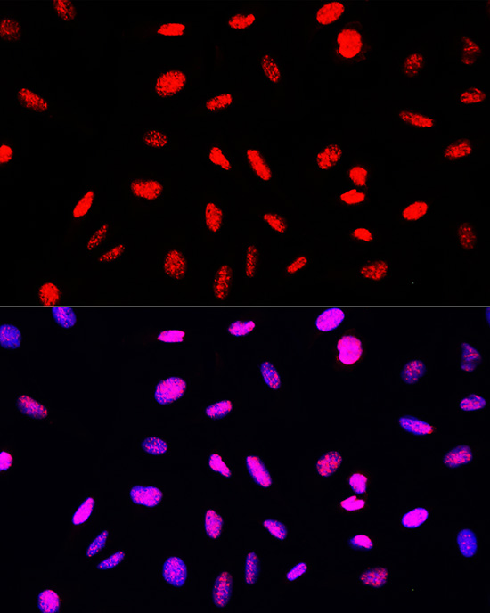
Immunofluorescence analysis of C6 cellsusing Phospho-Histone H3-S10/T11 RabbitpAbat dilution of 100 (40x lens).Secondary antibody: Cy3-conjugated Goatanti-Rabbit IgG (H+L) at 1:500dilution. Blue: DAPI for nuclear staining.ICC/IF
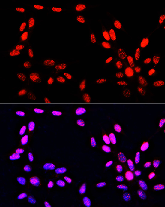
Immunofluorescence analysis of NIH-3T3cells using Phospho-Histone H3-S10/T11Rabbit pAb at dilution of 100 (40xlens). Secondary antibody: Cy3-conjugatedGoat anti-Rabbit IgG (H+L) at 1:500dilution. Blue: DAPI for nuclear staining.ICC/IF
Immunofluorescence analysis of U-2 OS cellsusing Phospho-Histone H3-S10/T11 RabbitpAb at dilution of 100 (40x lens).Secondary antibody: Cy3-conjugated Goatanti-Rabbit IgG (H+L) at 1:500dilution. Blue: DAPI for nuclear staining.IHC
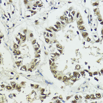
Immunohistochemistry analysis of paraffinembedded Human lung cancer usingPhospho-Histone H3-S10/T11 Rabbit pAb at dilution of 1:100 (40x lens).Microwave antigen retrieval performed with0.01M Tris/EDTA Buffer (pH 9.0) prior to IHCstaining.IHC
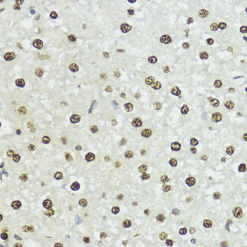
Immunohistochemistry analysis of paraffinembedded Rat liver using Phospho-HistoneH3-S10/T11 Rabbit pAb at dilutionof 1:100 (40x lens). Microwave antigenretrieval performed with 0.01M Tris/EDTABuffer (pH 9.0) prior to IHC staining.IHC
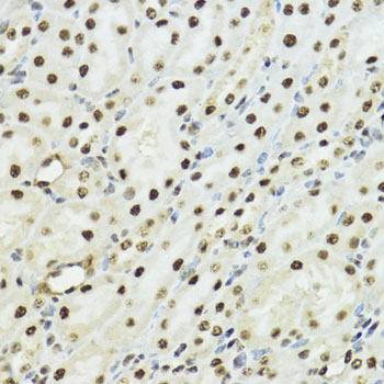
Immunohistochemistry analysis of paraffinembedded Mouse kidney using PhosphoHistone H3-S10/T11 Rabbit pAb at dilution of 1:100 (40x lens). Microwaveantigen retrieval performed with 0.01MTris/EDTA Buffer (pH 9.0) prior to IHCstaining.| Product Name | Phospho-Histone H3-S10/T11 Rabbit pAb |
|---|---|
| Antibody Type | Primary Antibodies |
| Immunogen | Synthetic peptide. This information is considered to be commercially sensitive. |
| Modification | P-S10/T11 |
| Clonality | Polyclonal |
|---|---|
| Isotype | IgG |
| Host Species | Rabbit |
| Tested Applications | ICC/IFIHCWB |
| WB:1:500-1:2000 ICC/IF:1:50-1:200 IHC:1:50-1:200 |
|
| Species Reactivity | HumanMouseOtherRat |
| Concentration | 1mg/ml |
| Purification | Affinity purified |
| Gene Symbol | H3-4 |
|---|---|
| Gene Synonyms | H3t H3.4 H3/g H3FT H3C16 HIST3H3 |
| Gene Full Name | H3.4 histone, cluster member |
| Gene Summary | Histones are basic nuclear proteins that are responsible for the nucleosome structure of the chromosomal fiber in eukaryotes. Nucleosomes consist of approximately 146 bp of DNA wrapped around a histone octamer composed of pairs of each of the four core histones (H2A, H2B, H3, and H4). The chromatin fiber is further compacted through the interaction of a linker histone, H1, with the DNA between the nucleosomes to form higher order chromatin structures. This gene is intronless and encodes a replication-dependent histone that is a member of the histone H3 family. Transcripts from this gene lack polyA tails; instead, they contain a palindromic termination element. This gene is located separately from the other H3 genes that are in the histone gene cluster on chromosome 6p22-p21.3. [provided by RefSeq, Aug 2015] |
| Alternative Names | H3/A H3C2 H3C3 H3C4 H3C6 H3C7 H3C8 H3FA H3C10 H3C11 H3C12 HIST1H3A Phospho-Histone H3-S10/T11 |
| Molecular Weight(MW) | 15kDa(Observed band size:17kDa) |
| Cellular Localization | Chromosome, Nucleus. |
WB
Western blot analysis of lysates from NIH/3T3 cells, using Phospho-Histone H3-S10/T11 Rabbit pAb at 1:1000 dilution. HeLa,NIH/3T3 and C6 cellls were treated with nocodazole (50 ng/mL) at 37℃for 20 hours.Secondary antibody: HRP-conjugated Goat anti-Rabbit IgG (H+L) at 1:10000 dilution.Lysates/proteins: 25μg per lane.Blocking buffer: 3% BSA.Detection: ECL Basic Kit.Exposure time: 1s.
WB
Western blot analysis of various lysates using Phospho-Histone H3-S10/T11 Rabbit pAb at1:1000 dilution. Both NIH/3T3 cells and Hela cells were treated with CIP(20uL/400ul) at 37℃ for 1 hourand treated with nocodazole (50 ng/mL) at 37℃ for 20 hours.Secondary antibody: HRP-conjugated Goat anti-Rabbit IgG (H+L) at 1:10000 dilution.Lysates/proteins: 25μg per lane.Blocking buffer: 3% BSA.Detection: ECL Basic Kit.Exposure time: 1s.
ICC/IF
Immunofluorescence analysis of C6 cellsusing Phospho-Histone H3-S10/T11 RabbitpAbat dilution of 100 (40x lens).Secondary antibody: Cy3-conjugated Goatanti-Rabbit IgG (H+L) at 1:500dilution. Blue: DAPI for nuclear staining.
ICC/IF
Immunofluorescence analysis of NIH-3T3cells using Phospho-Histone H3-S10/T11Rabbit pAb at dilution of 100 (40xlens). Secondary antibody: Cy3-conjugatedGoat anti-Rabbit IgG (H+L) at 1:500dilution. Blue: DAPI for nuclear staining.
ICC/IF
Immunofluorescence analysis of U-2 OS cellsusing Phospho-Histone H3-S10/T11 RabbitpAb at dilution of 100 (40x lens).Secondary antibody: Cy3-conjugated Goatanti-Rabbit IgG (H+L) at 1:500dilution. Blue: DAPI for nuclear staining.
IHC
Immunohistochemistry analysis of paraffinembedded Human lung cancer usingPhospho-Histone H3-S10/T11 Rabbit pAb at dilution of 1:100 (40x lens).Microwave antigen retrieval performed with0.01M Tris/EDTA Buffer (pH 9.0) prior to IHCstaining.
IHC
Immunohistochemistry analysis of paraffinembedded Rat liver using Phospho-HistoneH3-S10/T11 Rabbit pAb at dilutionof 1:100 (40x lens). Microwave antigenretrieval performed with 0.01M Tris/EDTABuffer (pH 9.0) prior to IHC staining.
IHC
Immunohistochemistry analysis of paraffinembedded Mouse kidney using PhosphoHistone H3-S10/T11 Rabbit pAb at dilution of 1:100 (40x lens). Microwaveantigen retrieval performed with 0.01MTris/EDTA Buffer (pH 9.0) prior to IHCstaining.| Application Notes | WB:1:500-1:2000 ICC/IF:1:50-1:200 IHC:1:50-1:200 |
|---|
| Form | Liquid |
|---|---|
| Storage Instructions | Store at -20℃. Avoid freeze / thaw cycles. |
| Storage Buffer | PBS with 0.09% Sodium azide, 50% glycerol, pH7.3. |


