WB
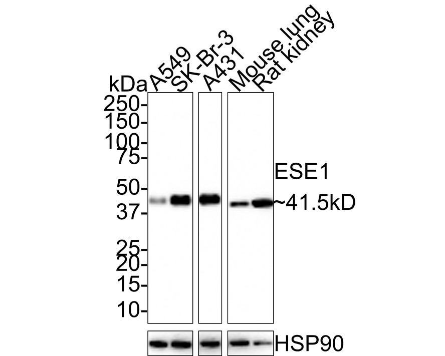
Western blot analysis of ESE1 on different lysates with Rabbit anti-ESE1 antibody at 1/1000 dilution. Lane 1: A549 cell lysate (10 µg/Lane), Lane 2: SK-Br-3 cell lysate (10 µg/Lane), Lane 3: A431 cell lysate (10 µg/Lane), Lane 4: Mouse lung tissue lysate (20 µg/Lane), Lane 5: Rat kidney tissue lysate (20 µg/Lane), Exposure time: 40 seconds; 4-20% SDS-PAGE gel. Proteins were transferred to a PVDF membrane and blocked with 5% NFDM/TBST for 1 hour at room temperature. The primary antibody at 1/1000 dilution was used in 5% NFDM/TBST at room temperature for 2 hours. Goat Anti-Rabbit IgG - HRP Secondary Antibody at 1:100000 dilution was used for 1 hour at room temperature.FC
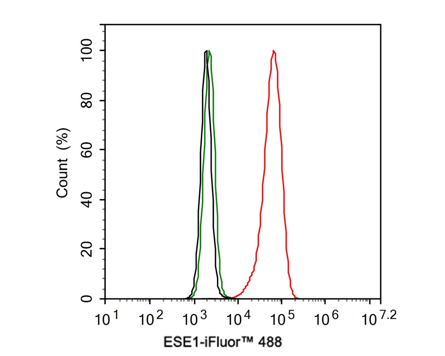
Flow cytometric analysis of A431 cells labeling ESE1. Cells were fixed and permeabilized. Then stained with the primary antibody (1ug/ml) (red) compared with Rabbit IgG Isotype Control (green). After incubation of the primary antibody at +4℃ for an hour, the cells were stained with a iFluor™ 488 conjugate-Goat anti-Rabbit IgG Secondary antibody at 1/1000 dilution for 30 minutes at +4℃. Unlabelled sample was used as a control (cells without incubation with primary antibody; black).| Product Name | ESE1 Recombinant Rabbit Monoclonal Antibody |
|---|---|
| Antibody Type | Primary Antibodies |
| Product description |
|
| Immunogen | Recombinant protein within Human ESE1 aa 1-371 / 371. |
| Clonality | monoclonal |
|---|---|
| Isotype | IgG |
| Host Species | Rabbit |
| Tested Applications | FCWB |
| WB:1:1000 FC:1:500-1:1000 |
|
| Species Reactivity | HumanMouseRat |
| Concentration | 1mg/ml |
| Purification | Protein A |
| Gene Symbol | ELF3 |
|---|---|
| Gene Synonyms | ERT ESX EPR-1 ESE-1 |
| Gene Full Name | E74 like ETS transcription factor 3 |
| Gene Summary | Enables DNA-binding transcription activator activity, RNA polymerase II-specific and sequence-specific double-stranded DNA binding activity. Involved in inflammatory response; negative regulation of DNA-templated transcription; and positive regulation of transcription by RNA polymerase II. Located in Golgi apparatus; cytosol; and nucleoplasm. [provided by Alliance of Genome Resources, Jul 2025] |
| Alternative Names | E74 like factor 3 antibody E74 like factor 3 ets domain transcription factor antibody E74 like factor 3 ets domain transcription factor epithelial specific antibody E74 like factor 3 ETS domain transcription factor serine box epithelial specific antibody E74-like factor 3 antibody Elf3 antibody ELF3_HUMAN antibody Epithelial restricted with serine box antibody Epithelial-restricted with serine box antibody Epithelium restricted Ets protein ESX antibody E74 like factor 3 antibody E74 like factor 3 ets domain transcription factor antibody E74 like factor 3 ets domain transcription factor epithelial specific antibody E74 like factor 3 ETS domain transcription factor serine box epithelial specific antibody E74-like factor 3 antibody Elf3 antibody ELF3_HUMAN antibody Epithelial restricted with serine box antibody Epithelial-restricted with serine box antibody Epithelium restricted Ets protein ESX antibody Epithelium specific Ets factor 1 antibody Epithelium specific Ets transcription factor 1 antibody Epithelium-restricted Ets protein ESX antibody Epithelium-specific Ets transcription factor 1 antibody EPR 1 antibody EPR1 antibody ERT antibody ESE-1 antibody ESX antibody Ets domain transcription factor serine box antibody Ets domain transcription factor serine box epithelial specific antibody Ets transcription factor antibody ETS-related transcription factor Elf-3 antibody jen antibody MGC139318 antibody |
| Molecular Weight(MW) | 41.5kDa(Observed band size:41.5kDa) |
| Cellular Localization | Cytoplasm, Nucleus. |
WB
Western blot analysis of ESE1 on different lysates with Rabbit anti-ESE1 antibody at 1/1000 dilution. Lane 1: A549 cell lysate (10 µg/Lane), Lane 2: SK-Br-3 cell lysate (10 µg/Lane), Lane 3: A431 cell lysate (10 µg/Lane), Lane 4: Mouse lung tissue lysate (20 µg/Lane), Lane 5: Rat kidney tissue lysate (20 µg/Lane), Exposure time: 40 seconds; 4-20% SDS-PAGE gel. Proteins were transferred to a PVDF membrane and blocked with 5% NFDM/TBST for 1 hour at room temperature. The primary antibody at 1/1000 dilution was used in 5% NFDM/TBST at room temperature for 2 hours. Goat Anti-Rabbit IgG - HRP Secondary Antibody at 1:100000 dilution was used for 1 hour at room temperature.
FC
Flow cytometric analysis of A431 cells labeling ESE1. Cells were fixed and permeabilized. Then stained with the primary antibody (1ug/ml) (red) compared with Rabbit IgG Isotype Control (green). After incubation of the primary antibody at +4℃ for an hour, the cells were stained with a iFluor™ 488 conjugate-Goat anti-Rabbit IgG Secondary antibody at 1/1000 dilution for 30 minutes at +4℃. Unlabelled sample was used as a control (cells without incubation with primary antibody; black).| Application Notes | WB:1:1000 FC:1:500-1:1000 |
|---|
| Form | Liquid |
|---|---|
| Storage Instructions | Shipped at 4℃. Store at +4℃ short term (1-2 weeks). It is recommended to aliquot into single-use upon delivery. Store at -20℃ long term. |
| Storage Buffer | 1*TBS (pH7.4), 0.05% BSA, 40% Glycerol. Preservative: 0.05% Sodium Azide. |


