WB
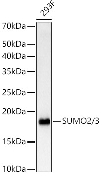
Western blot analysis of lysates from 293F cells, using SUMO2/3 Rabbit mAb at 1:1000 dilution.Secondary antibody: HRP-conjugated Goat anti-Rabbit IgG (H+L) at 1:10000 dilution.Lysates/proteins: 25μg per lane.Blocking buffer: 3% nonfat dry milk in TBST.Detection: ECL Basic Kit.Exposure time: 3s.WB
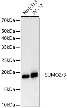
Western blot analysis of various lysates, using SUMO2/3 Rabbit mAb at 1:1000 dilution.Secondary antibody: HRP-conjugated Goat anti-Rabbit IgG (H+L) at 1:10000 dilution.Lysates/proteins: 25μg per lane.Blocking buffer: 3% nonfat dry milk in TBST.Detection: ECL Basic Kit.Exposure time: 3s.ICC/IF
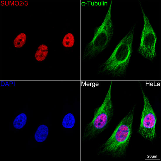
Confocal imaging of HeLa cells usingSUMO2/3 Rabbit mAb (dilution1:400)(Red). The cells were counterstainedwith α-Tubulin Mouse mAb (dilution1:400) (Green). DAPI was used for nuclearstaining (blue). Objective: 100x.| Product Name | SUMO2/3 Rabbit mAb |
|---|---|
| Antibody Type | Primary Antibodies |
| Immunogen | Recombinant protein (or fragment).This information is considered to be commercially sensitive. |
| Clonality | Monoclonal |
|---|---|
| Isotype | IgG |
| Host Species | Rabbit |
| Tested Applications | ICC/IFWB |
| WB:1:1000-1:6000 ICC/IF:1:400-1:1600 |
|
| Species Reactivity | HumanMouseRat |
| Concentration | 1mg/ml |
| Purification | Affinity purified |
| Gene Symbol | SUMO2 |
|---|---|
| Gene Synonyms | HSMT3 SMT3B SUMO3 Smt3A SMT3H2 |
| Gene Full Name | small ubiquitin like modifier 2 |
| Gene Summary | This gene encodes a protein that is a member of the SUMO (small ubiquitin-like modifier) protein family. It functions in a manner similar to ubiquitin in that it is bound to target proteins as part of a post-translational modification system. However, unlike ubiquitin which targets proteins for degradation, this protein is involved in a variety of cellular processes, such as nuclear transport, transcriptional regulation, apoptosis, and protein stability. It is not active until the last two amino acids of the carboxy-terminus have been cleaved off. Numerous pseudogenes have been reported for this gene. Alternate transcriptional splice variants, encoding different isoforms, have been characterized. [provided by RefSeq, Jul 2008] |
| Alternative Names | HSMT3 SMT3B SUMO3 Smt3A SMT3H2 SUMO2/3 |
| Molecular Weight(MW) | 11kDa(Observed band size:17kDa) |
| Cellular Localization | Nucleus, PML body. |
WB
Western blot analysis of lysates from 293F cells, using SUMO2/3 Rabbit mAb at 1:1000 dilution.Secondary antibody: HRP-conjugated Goat anti-Rabbit IgG (H+L) at 1:10000 dilution.Lysates/proteins: 25μg per lane.Blocking buffer: 3% nonfat dry milk in TBST.Detection: ECL Basic Kit.Exposure time: 3s.
WB
Western blot analysis of various lysates, using SUMO2/3 Rabbit mAb at 1:1000 dilution.Secondary antibody: HRP-conjugated Goat anti-Rabbit IgG (H+L) at 1:10000 dilution.Lysates/proteins: 25μg per lane.Blocking buffer: 3% nonfat dry milk in TBST.Detection: ECL Basic Kit.Exposure time: 3s.
ICC/IF
Confocal imaging of HeLa cells usingSUMO2/3 Rabbit mAb (dilution1:400)(Red). The cells were counterstainedwith α-Tubulin Mouse mAb (dilution1:400) (Green). DAPI was used for nuclearstaining (blue). Objective: 100x.| Application Notes | WB:1:1000-1:6000 ICC/IF:1:400-1:1600 |
|---|
| Form | Liquid |
|---|---|
| Storage Instructions | Store at -20℃. Avoid freeze / thaw cycles. |
| Storage Buffer | PBS with 0.09% Sodium azide, 0.05% BSA, 50% glycerol, pH7.3. |


