WB
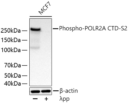
Western blot analysis of lysates from MCF7 cells using Phospho-POLR2A CTD-S2 Rabbit mAb at 1:5000 dilution incubated overnight at 4℃. MCF7 cells were treated with λpp (2 U/ul) at 30℃ for 1 hours.Secondary antibody: HRP-conjugated Goat anti-Rabbit IgG (H+L) at 1:10000 dilution.Lysates/proteins: 30 μg per lane.Blocking buffer: 3% nonfat dry milk in TBST.Detection: ECL Basic Kit.Exposure time: 20 s.WB
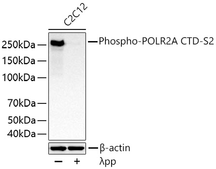
Western blot analysis of lysates from C2C12 cells using Phospho-POLR2A CTD-S2 Rabbit mAb at1:5000 dilution incubated overnight at 4℃. C2C12 cells were treated with λpp (2 U/ul) at 30℃ for 1 hours.Secondary antibody: HRP-conjugated Goat anti-Rabbit IgG (H+L) at 1:10000 dilution.Lysates/proteins: 30 μg per lane.Blocking buffer: 3% nonfat dry milk in TBST.Detection: ECL Basic Kit.Exposure time: 45 s.ICC/IF
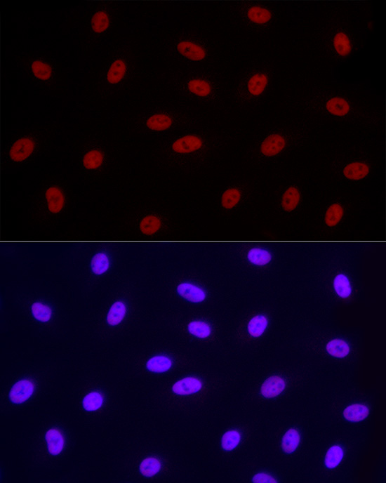
Immunofluorescence analysis of A-549 cellsusing Phospho-POLR2A CTD-S2 Rabbit mAb at dilution of 1:100 (40x lens).Secondary antibody: Cy3-conjugated Goatanti-Rabbit IgG (H+L) at 1:500dilution. Blue: DAPI for nuclear staining.ICC/IF
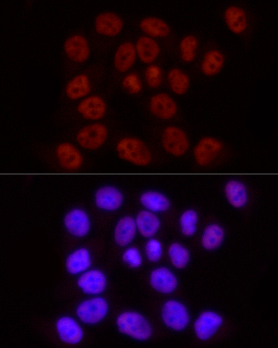
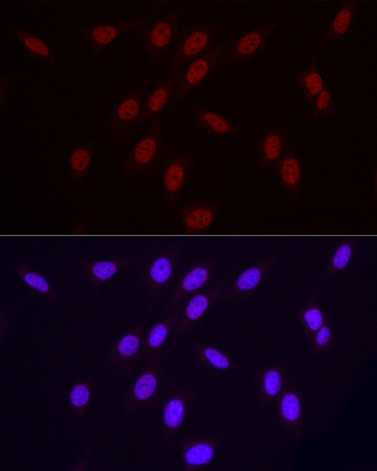
Immunofluorescence analysis of HeLa cellsusing Phospho-POLR2A CTD-S2 Rabbit mAb at dilution of 1:100 (40x lens).Secondary antibody: Cy3-conjugated Goatanti-Rabbit IgG (H+L) at 1:500dilution. Blue: DAPI for nuclear staining.ICC/IF
Immunofluorescence analysis of NIH/3T3cells using Phospho-POLR2A CTD-S2 RabbitmAb at dilution of 1:100 (40x lens).Secondary antibody: Cy3-conjugated Goatanti-Rabbit IgG (H+L) at 1:500dilution. Blue: DAPI for nuclear staining.ICC/IF
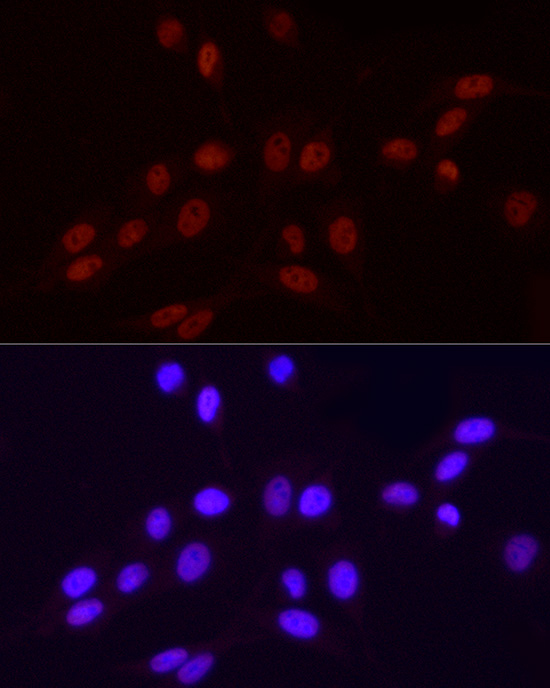
Immunofluorescence analysis of PC-12 cellsusing Phospho-POLR2A CTD-S2 Rabbit mAb at dilution of 1:100 (40x lens).Secondary antibody: Cy3-conjugated Goatanti-Rabbit IgG (H+L) at 1:500dilution. Blue: DAPI for nuclear staining.IHC
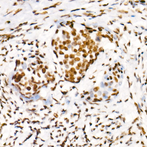
Immunohistochemistry analysis of paraffinembedded Human cervix cancer usingPhospho-POLR2A CTD-S2 Rabbit mAb at dilution of 1:200 (40x lens). Highpressure antigen retrieval performed with0.01M Citrate buffer (pH 6.0) prior to IHC staining.IHC
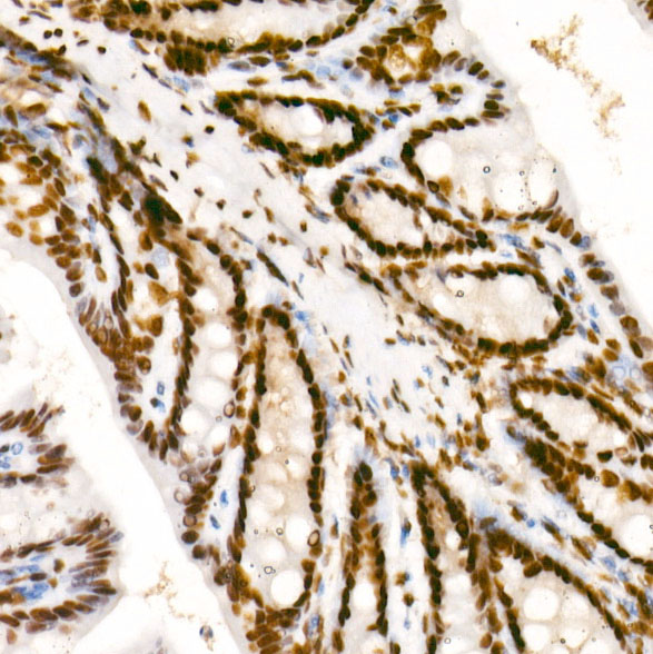
Immunohistochemistry analysis of paraffinembedded Mouse colon using PhosphoPOLR2A CTD-S2 Rabbit mAb at dilution of 1:200 (40x lens). High pressureantigen retrieval performed with 0.01MCitrate buffer (pH 6.0) prior to IHC staining.IHC
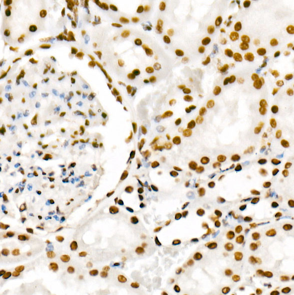
Immunohistochemistry analysis of paraffinembedded Rat kidney using PhosphoPOLR2A CTD-S2 Rabbit mAb at dilution of 1:200 (40x lens). High pressureantigen retrieval performed with 0.01MCitrate buffer (pH 6.0) prior to IHC staining.ChIP
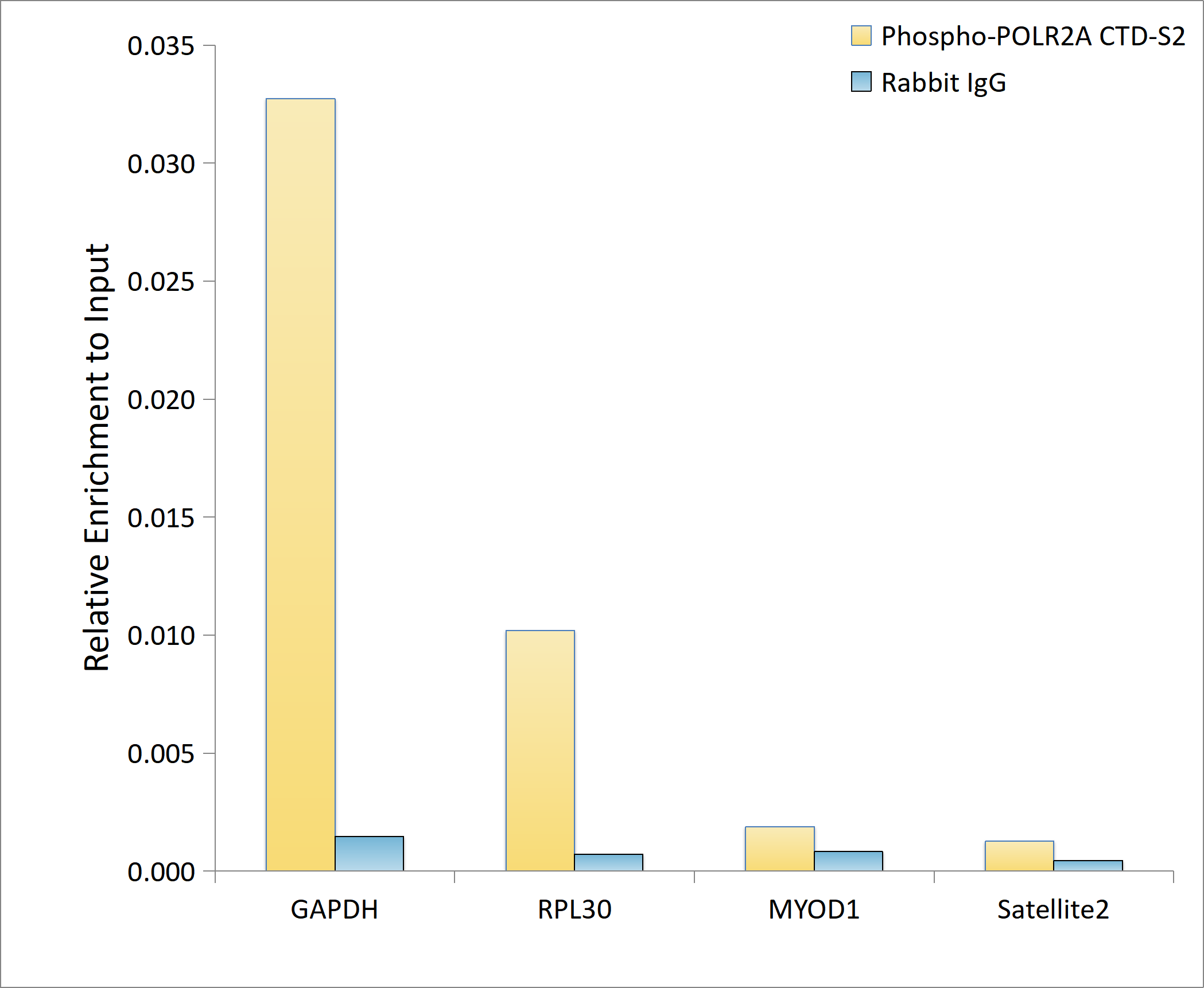
Chromatin immunoprecipitation analysis of extracts of 293F cells, using Phospho-POLR2A-S2 antibody and rabbit IgG.The amount of immunoprecipitated DNA was checked by quantitative PCR.Histogram was constructed by the ratios of the immunoprecipitated DNA to the input.CUT&Tag
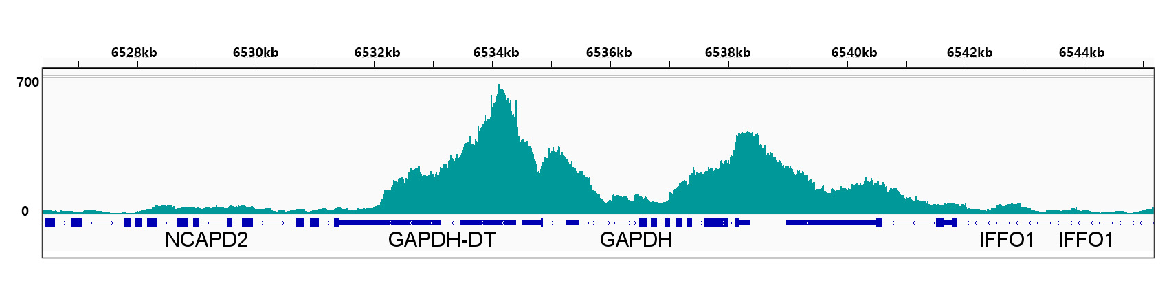
CUT&Tag was performed using the CUT&TagAssay Kit(pAG-Tn5) forIllumina from 10⁵ Hela cells with 1μg PhosphoPOLR2A CTD-S2 Rabbit mAb, alongwith a Goat Anti-Rabbit IgG(H+L). TheCUT&Tag results indicate the enrichmentpattern of Phospho-POLR2A CTD-S2 inrepresentative gene loci(GAPDH).CUT&Tag
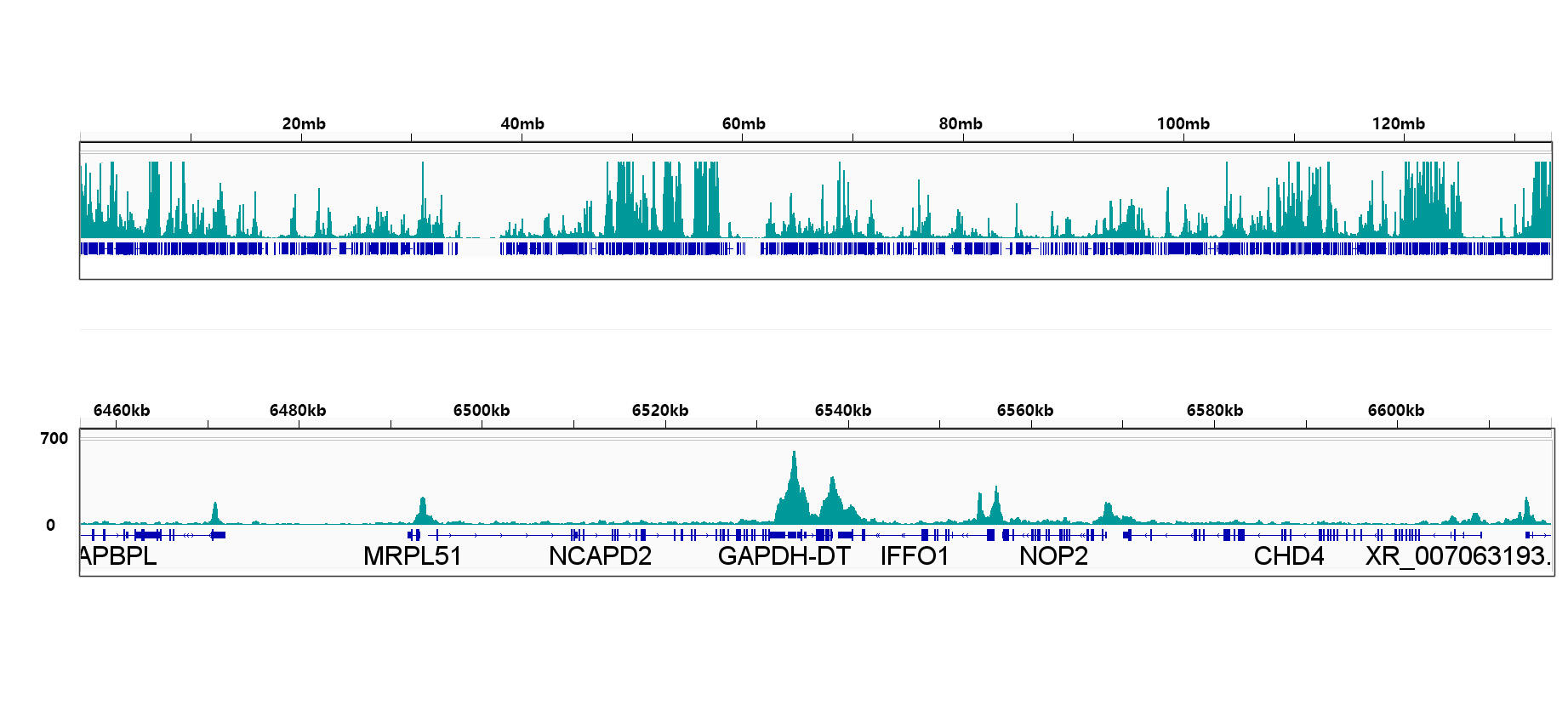
CUT&Tag was performed using the CUT&TagAssay Kit(pAG-Tn5) forIllumina from 10⁵ Hela cells with 1μg PhosphoPOLR2A CTD-S2 Rabbit mAb, alongwith a Goat Anti-Rabbit IgG(H+L). TheCUT&Tag results indicate the enrichmentpattern of Phospho-POLR2A CTD-S2 inrepresentative gene loci(GAPDH).| Product Name | Phospho-POLR2A CTD-S2 Rabbit mAb |
|---|---|
| Antibody Type | Primary Antibodies |
| Immunogen | Synthetic peptide. This information is considered to be commercially sensitive. |
| Modification | P-S2 |
| Clonality | Monoclonal |
|---|---|
| Isotype | IgG |
| Host Species | Rabbit |
| Tested Applications | ChIPICC/IFIHCWBCUT&Tag-seq |
| WB:1:3000-1:10000 ICC/IF:1:50-1:200 IHC:1:50-1:200 ChIP:5μg antibody for 10μg-15μg of Chromatin CUT&Tag:10⁵ cells /1 μg |
|
| Species Reactivity | HumanMouseRat |
| Concentration | 1mg/ml |
| Purification | Affinity purified |
| Gene Symbol | POLR2A |
|---|---|
| Gene Synonyms | RPB1 RPO2 POLR2 POLRA RPBh1 RPOL2 NEDHIB RpIILS hsRPB1 hRPB220 |
| Gene Full Name | RNA polymerase II subunit A |
| Gene Summary | This gene encodes the largest subunit of RNA polymerase II, the polymerase responsible for synthesizing messenger RNA in eukaryotes. The product of this gene contains a carboxy terminal domain composed of heptapeptide repeats that are essential for polymerase activity. These repeats contain serine and threonine residues that are phosphorylated in actively transcribing RNA polymerase. In addition, this subunit, in combination with several other polymerase subunits, forms the DNA binding domain of the polymerase, a groove in which the DNA template is transcribed into RNA. [provided by RefSeq, Jul 2008] |
| Alternative Names | RPB1 RPO2 POLR2 POLRA RPBh1 RPOL2 NEDHIB RpIILS hsRPB1 hRPB220 Phospho-POLR2A CTD-S2 |
| Molecular Weight(MW) | 217kDa(Observed band size:270kDa) |
| Cellular Localization | Nucleus. |
WB
Western blot analysis of lysates from MCF7 cells using Phospho-POLR2A CTD-S2 Rabbit mAb at 1:5000 dilution incubated overnight at 4℃. MCF7 cells were treated with λpp (2 U/ul) at 30℃ for 1 hours.Secondary antibody: HRP-conjugated Goat anti-Rabbit IgG (H+L) at 1:10000 dilution.Lysates/proteins: 30 μg per lane.Blocking buffer: 3% nonfat dry milk in TBST.Detection: ECL Basic Kit.Exposure time: 20 s.
WB
Western blot analysis of lysates from C2C12 cells using Phospho-POLR2A CTD-S2 Rabbit mAb at1:5000 dilution incubated overnight at 4℃. C2C12 cells were treated with λpp (2 U/ul) at 30℃ for 1 hours.Secondary antibody: HRP-conjugated Goat anti-Rabbit IgG (H+L) at 1:10000 dilution.Lysates/proteins: 30 μg per lane.Blocking buffer: 3% nonfat dry milk in TBST.Detection: ECL Basic Kit.Exposure time: 45 s.
ICC/IF
Immunofluorescence analysis of A-549 cellsusing Phospho-POLR2A CTD-S2 Rabbit mAb at dilution of 1:100 (40x lens).Secondary antibody: Cy3-conjugated Goatanti-Rabbit IgG (H+L) at 1:500dilution. Blue: DAPI for nuclear staining.
ICC/IF
Immunofluorescence analysis of HeLa cellsusing Phospho-POLR2A CTD-S2 Rabbit mAb at dilution of 1:100 (40x lens).Secondary antibody: Cy3-conjugated Goatanti-Rabbit IgG (H+L) at 1:500dilution. Blue: DAPI for nuclear staining.
ICC/IF
Immunofluorescence analysis of NIH/3T3cells using Phospho-POLR2A CTD-S2 RabbitmAb at dilution of 1:100 (40x lens).Secondary antibody: Cy3-conjugated Goatanti-Rabbit IgG (H+L) at 1:500dilution. Blue: DAPI for nuclear staining.
ICC/IF
Immunofluorescence analysis of PC-12 cellsusing Phospho-POLR2A CTD-S2 Rabbit mAb at dilution of 1:100 (40x lens).Secondary antibody: Cy3-conjugated Goatanti-Rabbit IgG (H+L) at 1:500dilution. Blue: DAPI for nuclear staining.
IHC
Immunohistochemistry analysis of paraffinembedded Human cervix cancer usingPhospho-POLR2A CTD-S2 Rabbit mAb at dilution of 1:200 (40x lens). Highpressure antigen retrieval performed with0.01M Citrate buffer (pH 6.0) prior to IHC staining.
IHC
Immunohistochemistry analysis of paraffinembedded Mouse colon using PhosphoPOLR2A CTD-S2 Rabbit mAb at dilution of 1:200 (40x lens). High pressureantigen retrieval performed with 0.01MCitrate buffer (pH 6.0) prior to IHC staining.
IHC
Immunohistochemistry analysis of paraffinembedded Rat kidney using PhosphoPOLR2A CTD-S2 Rabbit mAb at dilution of 1:200 (40x lens). High pressureantigen retrieval performed with 0.01MCitrate buffer (pH 6.0) prior to IHC staining.
ChIP
Chromatin immunoprecipitation analysis of extracts of 293F cells, using Phospho-POLR2A-S2 antibody and rabbit IgG.The amount of immunoprecipitated DNA was checked by quantitative PCR.Histogram was constructed by the ratios of the immunoprecipitated DNA to the input.
CUT&Tag
CUT&Tag was performed using the CUT&TagAssay Kit(pAG-Tn5) forIllumina from 10⁵ Hela cells with 1μg PhosphoPOLR2A CTD-S2 Rabbit mAb, alongwith a Goat Anti-Rabbit IgG(H+L). TheCUT&Tag results indicate the enrichmentpattern of Phospho-POLR2A CTD-S2 inrepresentative gene loci(GAPDH).
CUT&Tag
CUT&Tag was performed using the CUT&TagAssay Kit(pAG-Tn5) forIllumina from 10⁵ Hela cells with 1μg PhosphoPOLR2A CTD-S2 Rabbit mAb, alongwith a Goat Anti-Rabbit IgG(H+L). TheCUT&Tag results indicate the enrichmentpattern of Phospho-POLR2A CTD-S2 inrepresentative gene loci(GAPDH).| Application Notes | WB:1:3000-1:10000 ICC/IF:1:50-1:200 IHC:1:50-1:200 ChIP:5μg antibody for 10μg-15μg of Chromatin CUT&Tag:10⁵ cells /1 μg |
|---|
| Form | Liquid |
|---|---|
| Storage Instructions | Store at -20℃. Avoid freeze / thaw cycles. |
| Storage Buffer | PBS containing 50% glycerol and 0.05% BSA, preserved with proclin300 or sodium azide (as specified on the Certificate of Analysis), pH 7.3. |


