WB
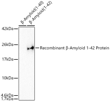
Western blot analysis of Recombinant β-Amyloid (1-40) protein and Recombinant β-Amyloid (1-42) proteinusing β-Amyloid(1-42) Rabbit mAb at 1:5000 dilution incubated overnight at 4℃.Secondary antibody: HRP-conjugated Goat anti-Rabbit IgG (H+L) at 1:10000 dilution.Lysates/proteins: 1 μg per lane.Blocking buffer: 3% nonfat dry milk in TBST.Detection: ECL Basic Kit.Negative control (NC): Recombinant β-Amyloid (1-40) protein.Exposure time: 90s.IHC
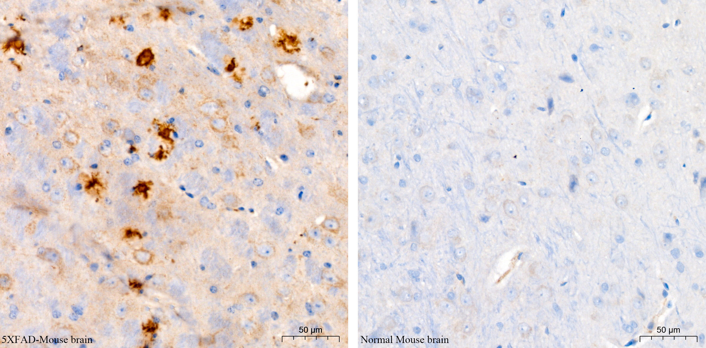
Immunohistochemistry analysis of paraffinembedded 5XFAD Mouse brain (model forAlzheimer disease, Left) and BALB/c Mousebrain (normal, Right) using β-Amyloid(1-42)Rabbit mAb at a dilution of 1:200(40x lens). High pressure antigen retrievalperformed with 0.01M Citrate buffer (pH 6.0)prior to IHC staining.IF-P
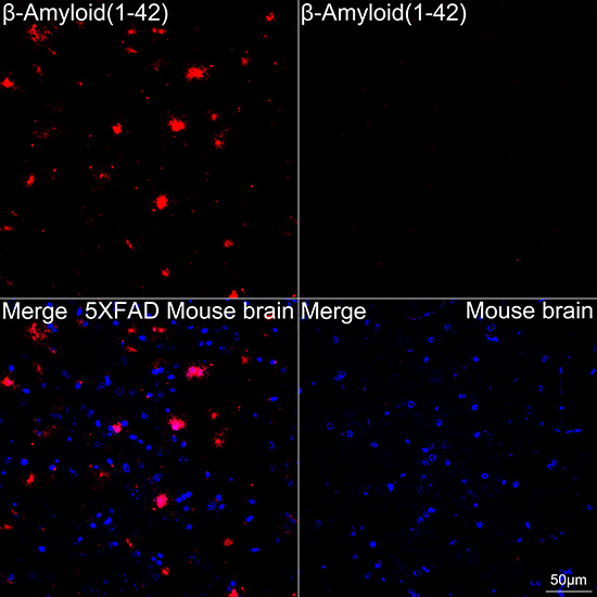
Confocal imaging of paraffin-embedded5XFAD Mouse brain (model for Alzheimerdisease, left) and BALB/c Mouse brain(normal, right) tissue using β-Amyloid(1-42)Rabbit mAb (dilution 1:200)followed by a further incubation with Cy3Goat Anti-Rabbit IgG (H+L) (dilution1:500) (Red). DAPI was used for nuclearstaining (Blue). High pressure antigenretrieval performed with 0.01M CitrateBuffer(pH 6.0) prior to IF staining. Objective:40x.DB
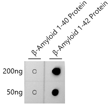
Dot-blot analysis of β-Amyloid 1-40 and βAmyloid 1-42 proteins using β-Amyloid(1-42)Rabbit mAb at 1:1000 dilution.| Product Name | β-Amyloid(1-42) Rabbit mAb |
|---|---|
| Antibody Type | Primary Antibodies |
| Immunogen | Synthetic peptide. This information is considered to be commercially sensitive. |
| Clonality | Monoclonal |
|---|---|
| Isotype | IgG |
| Host Species | Rabbit |
| Tested Applications | DBIF-PIHCWB |
| WB:1:1000-1:5000 IF-P:1:200-1:800 IHC:1:200-1:800 DB:1:500 - 1:1000 |
|
| Species Reactivity | Mouse |
| Concentration | 1mg/ml |
| Purification | Affinity purified |
| Gene Symbol | APP |
|---|---|
| Gene Synonyms | AAA AD1 PN2 ABPP APPI CVAP ABETA PN-II preA4 CTFgamma alpha-sAPP |
| Gene Full Name | amyloid beta precursor protein |
| Gene Summary | This gene encodes a cell surface receptor and transmembrane precursor protein that is cleaved by secretases to form a number of peptides. Some of these peptides are secreted and can bind to the acetyltransferase complex APBB1/TIP60 to promote transcriptional activation, while others form the protein basis of the amyloid plaques found in the brains of patients with Alzheimer disease. In addition, two of the peptides are antimicrobial peptides, having been shown to have bacteriocidal and antifungal activities. Mutations in this gene have been implicated in autosomal dominant Alzheimer disease and cerebroarterial amyloidosis (cerebral amyloid angiopathy). Multiple transcript variants encoding several different isoforms have been found for this gene. [provided by RefSeq, Aug 2014] |
| Alternative Names | AAA AD1 PN2 ABPP APPI CVAP ABETA PN-II preA4 CTFgamma alpha-sAPP β-Amyloid(1-42) |
| Molecular Weight(MW) | 4kDa(Observed band size:21kDa) |
| Cellular Localization | Membrane, Single-pass type I membrane protein, clathrin-coated pit. |
WB
Western blot analysis of Recombinant β-Amyloid (1-40) protein and Recombinant β-Amyloid (1-42) proteinusing β-Amyloid(1-42) Rabbit mAb at 1:5000 dilution incubated overnight at 4℃.Secondary antibody: HRP-conjugated Goat anti-Rabbit IgG (H+L) at 1:10000 dilution.Lysates/proteins: 1 μg per lane.Blocking buffer: 3% nonfat dry milk in TBST.Detection: ECL Basic Kit.Negative control (NC): Recombinant β-Amyloid (1-40) protein.Exposure time: 90s.
IHC
Immunohistochemistry analysis of paraffinembedded 5XFAD Mouse brain (model forAlzheimer disease, Left) and BALB/c Mousebrain (normal, Right) using β-Amyloid(1-42)Rabbit mAb at a dilution of 1:200(40x lens). High pressure antigen retrievalperformed with 0.01M Citrate buffer (pH 6.0)prior to IHC staining.
IF-P
Confocal imaging of paraffin-embedded5XFAD Mouse brain (model for Alzheimerdisease, left) and BALB/c Mouse brain(normal, right) tissue using β-Amyloid(1-42)Rabbit mAb (dilution 1:200)followed by a further incubation with Cy3Goat Anti-Rabbit IgG (H+L) (dilution1:500) (Red). DAPI was used for nuclearstaining (Blue). High pressure antigenretrieval performed with 0.01M CitrateBuffer(pH 6.0) prior to IF staining. Objective:40x.
DB
Dot-blot analysis of β-Amyloid 1-40 and βAmyloid 1-42 proteins using β-Amyloid(1-42)Rabbit mAb at 1:1000 dilution.| Application Notes | WB:1:1000-1:5000 IF-P:1:200-1:800 IHC:1:200-1:800 DB:1:500 - 1:1000 |
|---|
| Form | Liquid |
|---|---|
| Storage Instructions | Store at -20℃. Avoid freeze / thaw cycles. |
| Storage Buffer | PBS containing 50% glycerol and 0.05% BSA, preserved with proclin300 or sodium azide (as specified on the Certificate of Analysis), pH 7.3. |


