WB
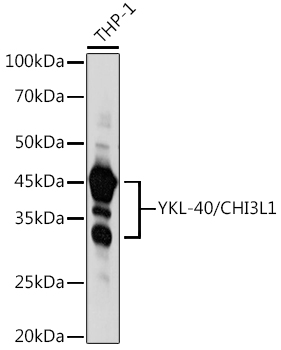
Western blot analysis of lysates from THP-1 cells, using YKL-40/CHI3L1 Rabbit pAb at 1:1000dilution.Secondary antibody: HRP-conjugated Goat anti-Rabbit IgG (H+L) at 1:10000 dilution.Lysates/proteins: 25μg per lane.Blocking buffer: 3% nonfat dry milk in TBST.Detection: ECL Basic Kit.Exposure time: 30s.ICC/IF
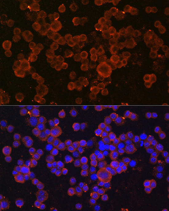
Immunofluorescence analysis of RAW264.7cells using YKL-40/CHI3L1 Rabbit pAb at dilution of 1:100 (40x lens).Secondary antibody: Cy3-conjugated Goatanti-Rabbit IgG (H+L) at 1:500dilution. Blue: DAPI for nuclear staining.ICC/IF
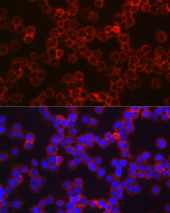
Immunofluorescence analysis of THP-1 cellsusing YKL-40/CHI3L1 Rabbit pAb at dilution of 1:100 (40x lens). Secondaryantibody: Cy3-conjugated Goat anti-RabbitIgG (H+L) at 1:500 dilution. Blue:DAPI for nuclear staining.IHC
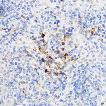
Immunohistochemistry analysis of paraffinembedded Rat spleen using YKL-40/CHI3L1Rabbit pAb at dilution of 1:100 (40xlens). Microwave antigen retrieval performedwith 0.01M PBS Buffer (pH 7.2) prior to IHC staining.IHC
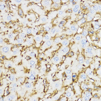
Immunohistochemistry analysis of paraffinembedded Mouse liver using YKL-40/CHI3L1Rabbit pAb at dilution of 1:100 (40xlens). Microwave antigen retrieval performedwith 0.01M PBS Buffer (pH 7.2) prior to IHC staining.| Product Name | YKL-40/CHI3L1 Rabbit pAb |
|---|---|
| Antibody Type | Primary Antibodies |
| Immunogen | This information is considered to be commercially sensitive. |
| Clonality | Polyclonal |
|---|---|
| Isotype | IgG |
| Host Species | Rabbit |
| Tested Applications | ICC/IFIHCWB |
| WB:1:500-1:5000 ICC/IF:1:50-1:200 IHC:1:50-1:200 |
|
| Species Reactivity | HumanMouseRat |
| Concentration | 1mg/ml |
| Purification | Affinity purified |
| Gene Symbol | CHI3L1 |
|---|---|
| Gene Synonyms | GP39 ASRT7 GP-39 YK-40 YKL40 CGP-39 YKL-40 YYL-40 HC-gp39 HCGP-3P hCGP-39 |
| Gene Full Name | chitinase 3 like 1 |
| Gene Summary | Chitinases catalyze the hydrolysis of chitin, which is an abundant glycopolymer found in insect exoskeletons and fungal cell walls. The glycoside hydrolase 18 family of chitinases includes eight human family members. This gene encodes a glycoprotein member of the glycosyl hydrolase 18 family. The protein lacks chitinase activity and is secreted by activated macrophages, chondrocytes, neutrophils and synovial cells. The protein is thought to play a role in the process of inflammation and tissue remodeling. [provided by RefSeq, Sep 2009] |
| Molecular Weight(MW) | 43kDa(Observed band size:30-45kDa) |
| Cellular Localization | Cytoplasm, Endoplasmic reticulum, Secreted, extracellular space, perinuclear region. |
WB
Western blot analysis of lysates from THP-1 cells, using YKL-40/CHI3L1 Rabbit pAb at 1:1000dilution.Secondary antibody: HRP-conjugated Goat anti-Rabbit IgG (H+L) at 1:10000 dilution.Lysates/proteins: 25μg per lane.Blocking buffer: 3% nonfat dry milk in TBST.Detection: ECL Basic Kit.Exposure time: 30s.
ICC/IF
Immunofluorescence analysis of RAW264.7cells using YKL-40/CHI3L1 Rabbit pAb at dilution of 1:100 (40x lens).Secondary antibody: Cy3-conjugated Goatanti-Rabbit IgG (H+L) at 1:500dilution. Blue: DAPI for nuclear staining.
ICC/IF
Immunofluorescence analysis of THP-1 cellsusing YKL-40/CHI3L1 Rabbit pAb at dilution of 1:100 (40x lens). Secondaryantibody: Cy3-conjugated Goat anti-RabbitIgG (H+L) at 1:500 dilution. Blue:DAPI for nuclear staining.
IHC
Immunohistochemistry analysis of paraffinembedded Rat spleen using YKL-40/CHI3L1Rabbit pAb at dilution of 1:100 (40xlens). Microwave antigen retrieval performedwith 0.01M PBS Buffer (pH 7.2) prior to IHC staining.
IHC
Immunohistochemistry analysis of paraffinembedded Mouse liver using YKL-40/CHI3L1Rabbit pAb at dilution of 1:100 (40xlens). Microwave antigen retrieval performedwith 0.01M PBS Buffer (pH 7.2) prior to IHC staining.| Application Notes | WB:1:500-1:5000 ICC/IF:1:50-1:200 IHC:1:50-1:200 |
|---|
| Form | Liquid |
|---|---|
| Storage Instructions | Store at -20℃. Avoid freeze / thaw cycles. |
| Storage Buffer | PBS containing 50% glycerol, preserved with proclin300 or sodium azide (as specified on the Certificate of Analysis), pH 7.3. |


