WB
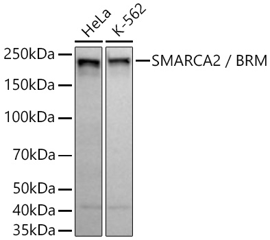
Western blot analysis of various lysates using SMARCA2 / BRM Rabbit mAb at 1:1000 dilutionincubated overnight at 4℃.Secondary antibody: HRP-conjugated Goat anti-Rabbit IgG (H+L) at 1:10000 dilution.Lysates/proteins: 25 μg per lane.Blocking buffer: 3% nonfat dry milk in TBST.Detection: ECL Basic Kit.Exposure time: 10 s.ICC/IF
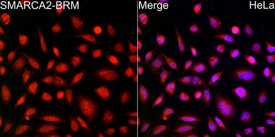
Immunofluorescence analysis of HeLa cellsusing SMARCA2 / BRM Rabbit mAb at dilution of 1:100 (40x lens). Secondaryantibody: Cy3-conjugated Goat anti-RabbitIgG (H+L) at 1:500 dilution. Blue:DAPI for nuclear staining.IHC
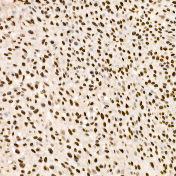
Immunohistochemistry analysis of paraffinembedded Human cervix cancer tissue usingSMARCA2 / BRM Rabbit mAb atdilution of 1:200 (40x lens). High pressureantigen retrieval performed with 0.01MCitrate Buffer (pH 6.0) prior to IHC staining.IP
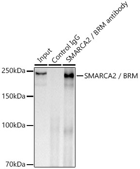
Immunoprecipitation analysis of 300 μg extracts of Hela cells using 3 μg SMARCA2 / BRM Rabbit mAb. Western blot was performed from the immunoprecipitate using SMARCA2 / BRM Rabbit mAb at a dilition of 1:5000.ChIP
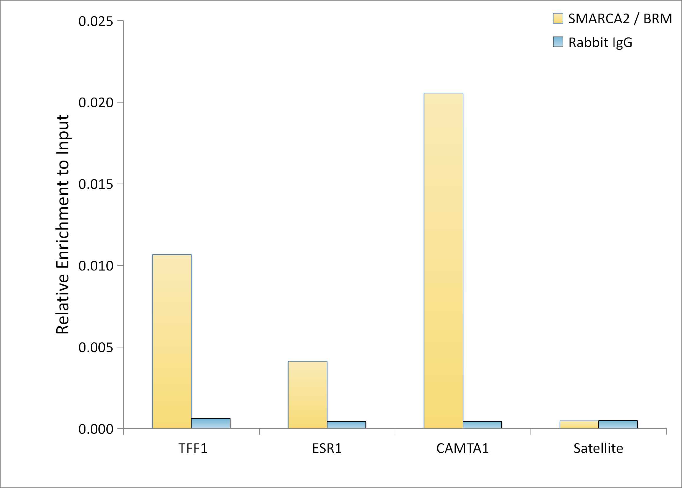
Chromatin immunoprecipitation was performed with 20 μg of cross-linked chromatin from MCF7 cellstreated with β-estradiol (10 nM, 45 min), using 5 μg of SMARCA2 / BRM Rabbit mAb and RabbitIgG isotype control. The enrichment of immunoprecipitated DNA at different genomic loci wasexamined by quantitative PCR. The histogram compares the ratio of the immunoprecipitated DNA to theinput at given loci.| Product Name | SMARCA2 / BRM Rabbit mAb |
|---|---|
| Antibody Type | Primary Antibodies |
| Immunogen | A synthetic peptide corresponding to a sequence within amino acids 201-300 of human SMARCA2 (NP_003061.3). |
| Clonality | Monoclonal |
|---|---|
| Isotype | IgG |
| Host Species | Rabbit |
| Tested Applications | ChIPICC/IFIHCIPWB |
| WB:1:500-1:2000 ICC/IF:1:50-1:200 IHC:1:200-1:800 ChIP: 5μg antibody for 10μg-20μg of Chromatin IP:0.5μg-4μg antibody for 200μg-400μg extracts of whole cells |
|
| Species Reactivity | Human |
| Concentration | 1mg/ml |
| Purification | Affinity purified |
| Gene Symbol | SMARCA2 |
|---|---|
| Gene Synonyms | BIS BRM SNF2 SWI2 hBRM NCBRS Sth1p BAF190 SNF2L2 SNF2LA hSNF2a SAMRCA2 |
| Gene Full Name | SWI/SNF related BAF chromatin remodeling complex subunit ATPase 2 |
| Gene Summary | The protein encoded by this gene is a member of the SWI/SNF family of proteins and is highly similar to the brahma protein of Drosophila. Members of this family have helicase and ATPase activities and are thought to regulate transcription of certain genes by altering the chromatin structure around those genes. The encoded protein is part of the large ATP-dependent chromatin remodeling complex SNF/SWI, which is required for transcriptional activation of genes normally repressed by chromatin. Alternatively spliced transcript variants encoding different isoforms have been found for this gene, which contains a trinucleotide repeat (CAG) length polymorphism. [provided by RefSeq, Jan 2014] |
| Alternative Names | BIS BRM SNF2 SWI2 hBRM NCBRS Sth1p BAF190 SNF2L2 SNF2LA hSNF2a SMARCA2 / BRM |
| Molecular Weight(MW) | 181kDa(Observed band size:200kDa) |
| Cellular Localization | nucleus. |
WB
Western blot analysis of various lysates using SMARCA2 / BRM Rabbit mAb at 1:1000 dilutionincubated overnight at 4℃.Secondary antibody: HRP-conjugated Goat anti-Rabbit IgG (H+L) at 1:10000 dilution.Lysates/proteins: 25 μg per lane.Blocking buffer: 3% nonfat dry milk in TBST.Detection: ECL Basic Kit.Exposure time: 10 s.
ICC/IF
Immunofluorescence analysis of HeLa cellsusing SMARCA2 / BRM Rabbit mAb at dilution of 1:100 (40x lens). Secondaryantibody: Cy3-conjugated Goat anti-RabbitIgG (H+L) at 1:500 dilution. Blue:DAPI for nuclear staining.
IHC
Immunohistochemistry analysis of paraffinembedded Human cervix cancer tissue usingSMARCA2 / BRM Rabbit mAb atdilution of 1:200 (40x lens). High pressureantigen retrieval performed with 0.01MCitrate Buffer (pH 6.0) prior to IHC staining.
IP
Immunoprecipitation analysis of 300 μg extracts of Hela cells using 3 μg SMARCA2 / BRM Rabbit mAb. Western blot was performed from the immunoprecipitate using SMARCA2 / BRM Rabbit mAb at a dilition of 1:5000.
ChIP
Chromatin immunoprecipitation was performed with 20 μg of cross-linked chromatin from MCF7 cellstreated with β-estradiol (10 nM, 45 min), using 5 μg of SMARCA2 / BRM Rabbit mAb and RabbitIgG isotype control. The enrichment of immunoprecipitated DNA at different genomic loci wasexamined by quantitative PCR. The histogram compares the ratio of the immunoprecipitated DNA to theinput at given loci.| Application Notes | WB:1:500-1:2000 ICC/IF:1:50-1:200 IHC:1:200-1:800 ChIP: 5μg antibody for 10μg-20μg of Chromatin IP:0.5μg-4μg antibody for 200μg-400μg extracts of whole cells |
|---|
| Form | Liquid |
|---|---|
| Storage Instructions | Store at -20℃. Avoid freeze / thaw cycles. |
| Storage Buffer | PBS containing 50% glycerol and 0.05% BSA, preserved with proclin300 or sodium azide (as specified on the Certificate of Analysis), pH 7.3. |


