WB
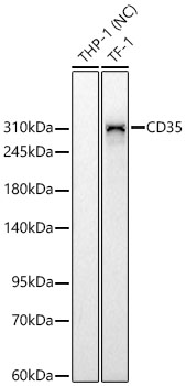
Western blot analysis of various lysates using CD35 Rabbit mAb at 1:1000 dilution.Secondary antibody: HRP-conjugated Goat anti-Rabbit IgG (H+L) at 1:10000 dilution.Lysates/proteins: 25 μg per lane.Blocking buffer: 3% nonfat dry milk in TBST.Detection: ECL Basic Kit.Negative control (NC): THP-1.Exposure time: 90s.ICC/IF
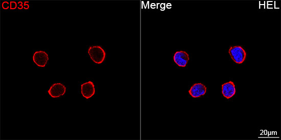
Confocal imaging of HEL cells using CD35Rabbit mAb (dilution 1:200)followed by a further incubation with Cy3Goat Anti-Rabbit IgG (H+L) (dilution1:500) (Red). DAPI was used for nuclearstaining (Blue). Objective: 100x.ICC/IF
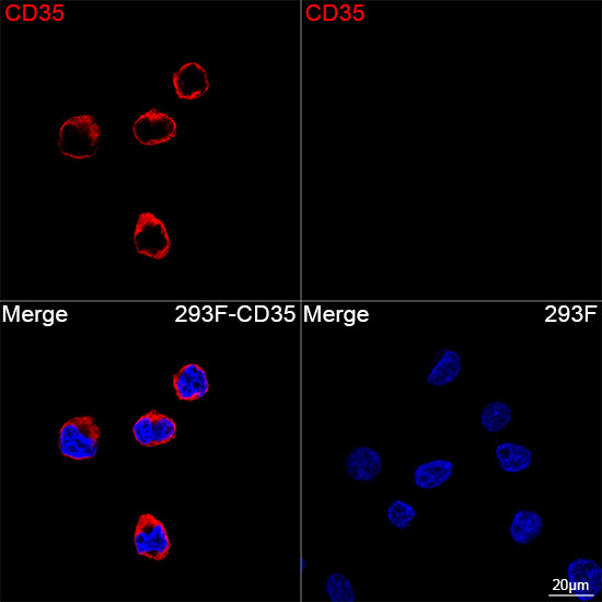
Confocal imaging of 293F cells transfectedwith CD35 using CD35 Rabbit mAb (dilution 1:200) followed by a furtherincubation with Cy3 Goat Anti-Rabbit IgG(H+L) (dilution 1:500) (Red). DAPIwas used for nuclear staining (Blue).Objective: 100x.IHC
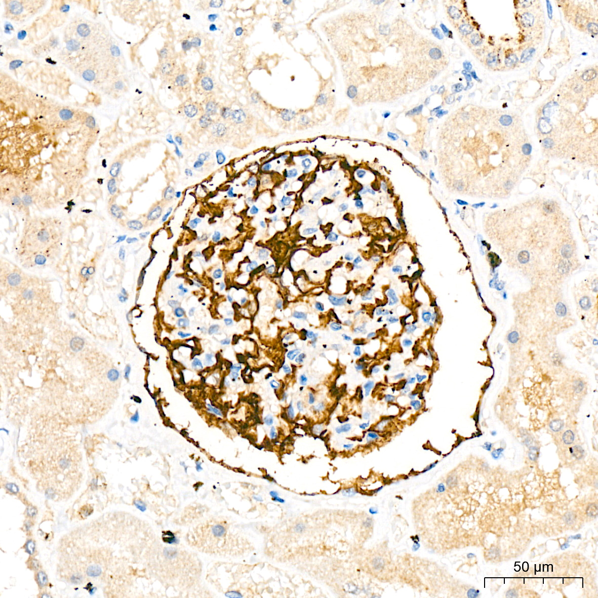
Immunohistochemistry analysis of paraffinembedded Human kidney tissue using CD35Rabbit mAb at a dilution of 1:200(40x lens). High pressure antigen retrievalwas performed with 0.01 M citrate buffer (pH6.0) prior to IHC staining.| Product Name | CD35 Rabbit mAb |
|---|---|
| Antibody Type | Primary Antibodies |
| Immunogen | Recombinant protein (or fragment).This information is considered to be commercially sensitive. |
| Clonality | Monoclonal |
|---|---|
| Isotype | IgG |
| Host Species | Rabbit |
| Tested Applications | ICC/IFIHCWB |
| WB:1:1000-1:2000 ICC/IF:1:200-1:2000 IHC:1:400-1:2000 |
|
| Species Reactivity | Human |
| Concentration | 1mg/ml |
| Purification | Affinity purified |
| Gene Symbol | CR1 |
|---|---|
| Gene Synonyms | KN C3BR C4BR CD35 |
| Gene Full Name | complement C3b/C4b receptor 1 (Knops blood group) |
| Gene Summary | This gene is a member of the receptors of complement activation (RCA) family and is located in the 'cluster RCA' region of chromosome 1. The genome is polymorphic at this locus with allele-specific splice variants encoding different isoforms, based on the presence/absence of long homologous repeats (LHRs). The gene encodes a monomeric single-pass type I membrane glycoprotein found on erythrocytes, leukocytes, glomerular podocytes, and splenic follicular dendritic cells. The Knops blood group system is a system of antigens located on this protein. The protein mediates cellular binding to particles and immune complexes that have activated complement. Decreases in expression of this protein and/or mutations in this gene have been associated with gallbladder carcinomas, mesangiocapillary glomerulonephritis, systemic lupus erythematosus, sarcoidosis and Alzheimer's disease. Mutations in this gene have also been associated with a reduction in Plasmodium falciparum rosetting, conferring protection against severe malaria. [provided by RefSeq, May 2020] |
| Alternative Names | KN C3BR C4BR CD35 |
| Molecular Weight(MW) | 224kDa(Observed band size:270kDa) |
| Cellular Localization | cell surface, cytoskeleton, extracellular exosome, extracellular space, plasma membrane, plasma membrane raft, Membrane, Single-pass type I membrane protein. |
WB
Western blot analysis of various lysates using CD35 Rabbit mAb at 1:1000 dilution.Secondary antibody: HRP-conjugated Goat anti-Rabbit IgG (H+L) at 1:10000 dilution.Lysates/proteins: 25 μg per lane.Blocking buffer: 3% nonfat dry milk in TBST.Detection: ECL Basic Kit.Negative control (NC): THP-1.Exposure time: 90s.
ICC/IF
Confocal imaging of HEL cells using CD35Rabbit mAb (dilution 1:200)followed by a further incubation with Cy3Goat Anti-Rabbit IgG (H+L) (dilution1:500) (Red). DAPI was used for nuclearstaining (Blue). Objective: 100x.
ICC/IF
Confocal imaging of 293F cells transfectedwith CD35 using CD35 Rabbit mAb (dilution 1:200) followed by a furtherincubation with Cy3 Goat Anti-Rabbit IgG(H+L) (dilution 1:500) (Red). DAPIwas used for nuclear staining (Blue).Objective: 100x.
IHC
Immunohistochemistry analysis of paraffinembedded Human kidney tissue using CD35Rabbit mAb at a dilution of 1:200(40x lens). High pressure antigen retrievalwas performed with 0.01 M citrate buffer (pH6.0) prior to IHC staining.| Application Notes | WB:1:1000-1:2000 ICC/IF:1:200-1:2000 IHC:1:400-1:2000 |
|---|
| Form | Liquid |
|---|---|
| Storage Instructions | Store at -20℃. Avoid freeze / thaw cycles. |
| Storage Buffer | PBS containing 50% glycerol and 0.05% BSA, preserved with proclin300 or sodium azide (as specified on the Certificate of Analysis), pH 7.3. |


