WB
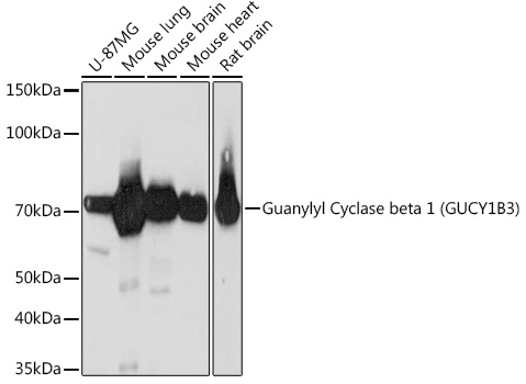
Western blot analysis of various lysates using Guanylyl Cyclase beta 1 (GUCY1B3) (GUCY1B3) Rabbit mAb at 1:1000 dilution.Secondary antibody: HRP-conjugated Goat anti-Rabbit IgG (H+L) at 1:10000 dilution.Lysates/proteins: 25μg per lane.Blocking buffer: 3% nonfat dry milk in TBST.Detection: ECL Basic Kit.Exposure time: 3min.ICC/IF
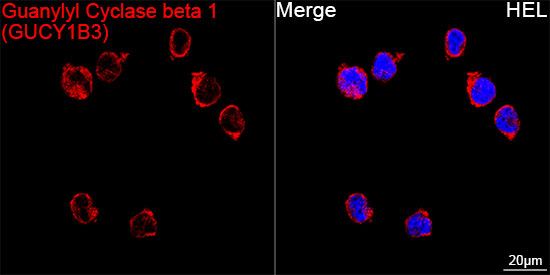
Confocal imaging of HEL cells using GuanylylCyclase beta 1 (GUCY1B3) Rabbit mAb(dilution 1:200) followed by a furtherincubation with Cy3 Goat Anti-Rabbit IgG(H+L) (dilution 1:500) (Red). DAPIwas used for nuclear staining (Blue).Objective: 100x.IHC
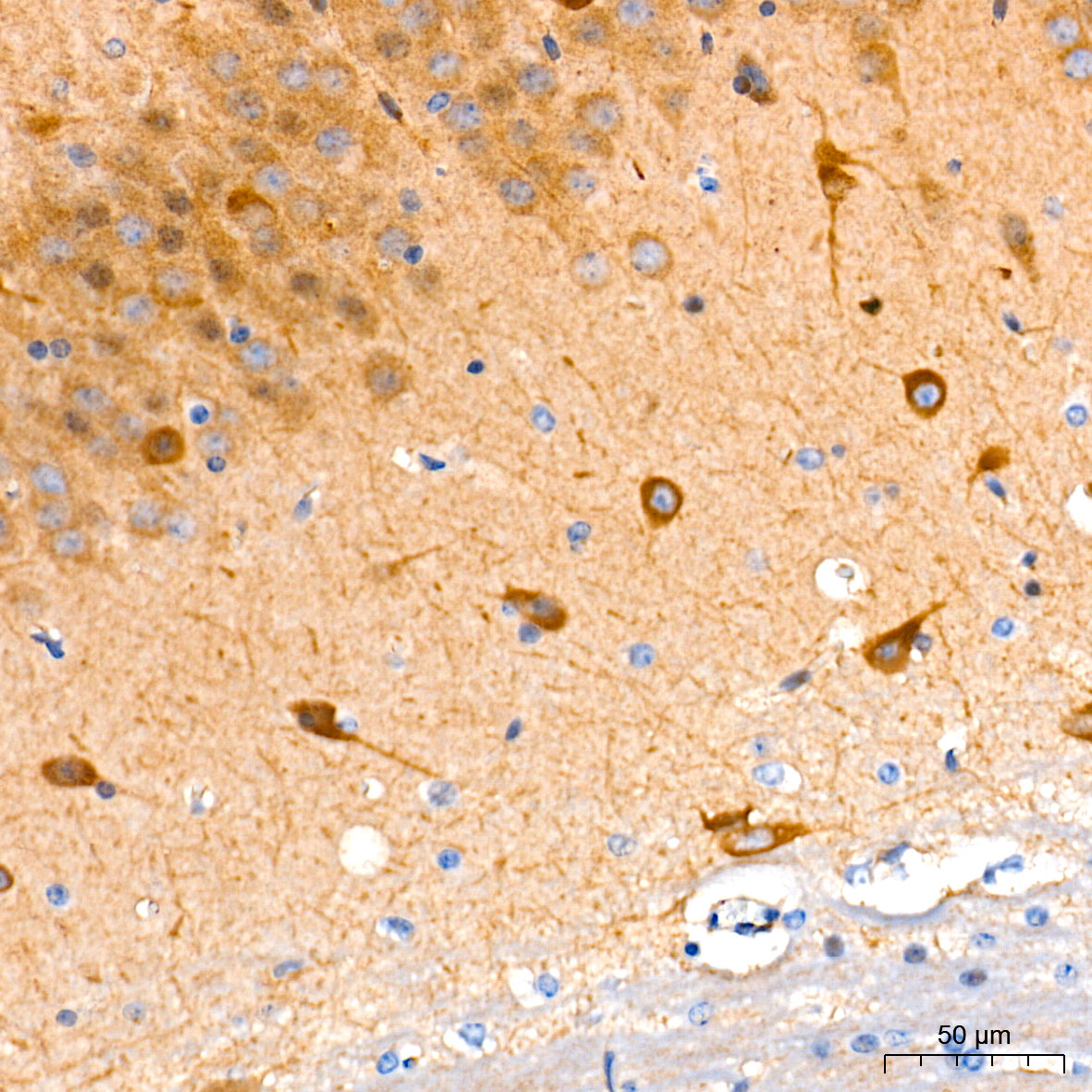
Immunohistochemistry analysis of paraffinembedded Mouse brain tissue using GuanylylCyclase beta 1 (GUCY1B3) Rabbit mAb at a dilution of 1:200 (40x lens).High pressure antigen retrieval performedwith 0.01M Tris-EDTA Buffer (pH 9.0) prior toIHC staining.IHC
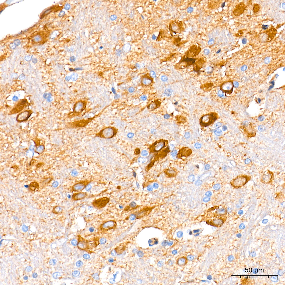
Immunohistochemistry analysis of paraffinembedded Rat brain tissue using GuanylylCyclase beta 1 (GUCY1B3) Rabbit mAb at a dilution of 1:200 (40x lens).High pressure antigen retrieval performedwith 0.01M Tris-EDTA Buffer (pH 9.0) prior to IHC staining.| Product Name | Guanylyl Cyclase beta 1 (GUCY1B3) Rabbit mAb |
|---|---|
| Antibody Type | Primary Antibodies |
| Immunogen | Synthetic peptide. This information is considered to be commercially sensitive. |
| Clonality | Monoclonal |
|---|---|
| Isotype | IgG |
| Host Species | Rabbit |
| Tested Applications | ICC/IFIHCWB |
| WB:1:1000-1:6000 IHC:1:200-1:800 ICC/IF:1:100-1:400 |
|
| Species Reactivity | HumanMouseRat |
| Concentration | 1mg/ml |
| Purification | Affinity purified |
| Gene Symbol | GUCY1B1 |
|---|---|
| Gene Synonyms | GUCB3 GC-SB3 GUC1B3 GUCSB3 GUCY1B3 GC-S-beta-1 |
| Gene Full Name | guanylate cyclase 1 soluble subunit beta 1 |
| Gene Summary | This gene encodes the beta subunit of the soluble guanylate cyclase (sGC), which catalyzes the conversion of GTP (guanosine triphosphate) to cGMP (cyclic guanosine monophosphate). The encoded protein contains an HNOX domain, which serves as a receptor for ligands such as nitric oxide, oxygen and nitrovasodilator drugs. Alternative splicing results in multiple transcript variants. [provided by RefSeq, May 2014] |
| Alternative Names | GUCB3 GC-SB3 GUC1B3 GUCSB3 GUCY1B3 GC-S-beta-1 Guanylyl Cyclase beta 1 (GUCY1B3) |
| Molecular Weight(MW) | 71kDa(Observed band size:71kDa) |
| Cellular Localization | Cytoplasm. |
WB
Western blot analysis of various lysates using Guanylyl Cyclase beta 1 (GUCY1B3) (GUCY1B3) Rabbit mAb at 1:1000 dilution.Secondary antibody: HRP-conjugated Goat anti-Rabbit IgG (H+L) at 1:10000 dilution.Lysates/proteins: 25μg per lane.Blocking buffer: 3% nonfat dry milk in TBST.Detection: ECL Basic Kit.Exposure time: 3min.
ICC/IF
Confocal imaging of HEL cells using GuanylylCyclase beta 1 (GUCY1B3) Rabbit mAb(dilution 1:200) followed by a furtherincubation with Cy3 Goat Anti-Rabbit IgG(H+L) (dilution 1:500) (Red). DAPIwas used for nuclear staining (Blue).Objective: 100x.
IHC
Immunohistochemistry analysis of paraffinembedded Mouse brain tissue using GuanylylCyclase beta 1 (GUCY1B3) Rabbit mAb at a dilution of 1:200 (40x lens).High pressure antigen retrieval performedwith 0.01M Tris-EDTA Buffer (pH 9.0) prior toIHC staining.
IHC
Immunohistochemistry analysis of paraffinembedded Rat brain tissue using GuanylylCyclase beta 1 (GUCY1B3) Rabbit mAb at a dilution of 1:200 (40x lens).High pressure antigen retrieval performedwith 0.01M Tris-EDTA Buffer (pH 9.0) prior to IHC staining.| Application Notes | WB:1:1000-1:6000 IHC:1:200-1:800 ICC/IF:1:100-1:400 |
|---|
| Form | Liquid |
|---|---|
| Storage Instructions | Store at -20℃. Avoid freeze / thaw cycles. |
| Storage Buffer | PBS containing 50% glycerol and 0.05% BSA, preserved with proclin300 or sodium azide (as specified on the Certificate of Analysis), pH 7.3. |


