WB
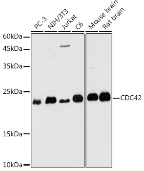
Western blot analysis of various lysates using CDC42 Rabbit pAb at 1:1000 dilution.Secondary antibody: HRP-conjugated Goat anti-Rabbit IgG (H+L) at 1:10000 dilution.Lysates/proteins: 25μg per lane.Blocking buffer: 3% nonfat dry milk in TBST.Detection: ECL Basic Kit.Exposure time: 180s.ICC/IF
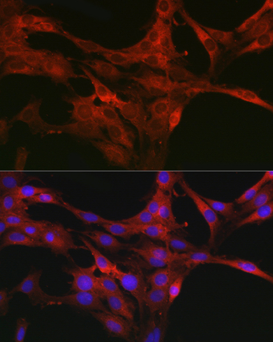
Immunofluorescence analysis of C6 cellsusing CDC42 Rabbit pAb at dilutionof 1:50 (40x lens). Secondary antibody: Cy3-conjugated Goat anti-Rabbit IgG (H+L) at 1:500 dilution. Blue: DAPI fornuclear staining.ICC/IF
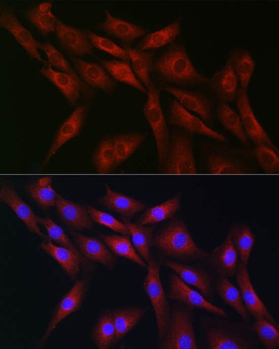
Immunofluorescence analysis of NIH-3T3cells using CDC42 Rabbit pAb a tdilution of 1:50 (40x lens). Secondaryantibody: Cy3-conjugated Goat anti-RabbitIgG (H+L) at 1:500 dilution. Blue:DAPI for nuclear staining.IHC
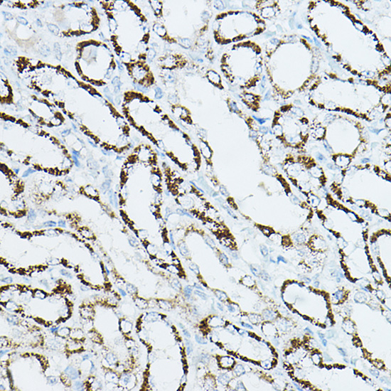
Immunohistochemistry analysis of paraffinembedded Rat kidney using CDC42 Rabbit pAb at dilution of 1:50 (40x lens).High pressure antigen retrieval performedwith 0.01M Citrate buffer (pH 6.0) prior toIHC staining.IHC
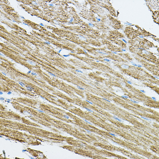
Immunohistochemistry analysis of paraffinembedded Mouse heart using CDC42 RabbitpAb at dilution of 1:50 (40x lens).High pressure antigen retrieval performedwith 0.01M Citrate buffer (pH 6.0) prior to IHC staining.| Product Name | CDC42 Rabbit pAb |
|---|---|
| Antibody Type | Primary Antibodies |
| Immunogen | Recombinant protein (or fragment).This information is considered to be commercially sensitive. |
| Clonality | Polyclonal |
|---|---|
| Isotype | IgG |
| Host Species | Rabbit |
| Tested Applications | ICC/IFIHCWB |
| WB:1:500-1:1000 IHC:1:50-1:200 ICC/IF:1:50-1:200 |
|
| Species Reactivity | HumanMouseRat |
| Concentration | 1mg/ml |
| Purification | Affinity purified |
| Gene Symbol | CDC42 |
|---|---|
| Gene Synonyms | TKS G25K CDC42Hs |
| Gene Full Name | cell division cycle 42 |
| Gene Summary | The protein encoded by this gene is a small GTPase of the Rho-subfamily, which regulates signaling pathways that control diverse cellular functions including cell morphology, migration, endocytosis and cell cycle progression. This protein is highly similar to Saccharomyces cerevisiae Cdc 42, and is able to complement the yeast cdc42-1 mutant. The product of oncogene Dbl was reported to specifically catalyze the dissociation of GDP from this protein. This protein could regulate actin polymerization through its direct binding to Neural Wiskott-Aldrich syndrome protein (N-WASP), which subsequently activates Arp2/3 complex. Alternative splicing of this gene results in multiple transcript variants. Pseudogenes of this gene have been identified on chromosomes 3, 4, 5, 7, 8 and 20. [provided by RefSeq, Apr 2013] |
| Alternative Names | TKS G25K CDC42Hs CDC42 |
| Molecular Weight(MW) | 21kDa(Observed band size:21kDa) |
| Cellular Localization | Cell membrane, Cytoplasm, Cytoplasmic side, Lipid-anchor, Midbody, centrosome, cytoskeleton, microtubule organizing center, spindle. |
WB
Western blot analysis of various lysates using CDC42 Rabbit pAb at 1:1000 dilution.Secondary antibody: HRP-conjugated Goat anti-Rabbit IgG (H+L) at 1:10000 dilution.Lysates/proteins: 25μg per lane.Blocking buffer: 3% nonfat dry milk in TBST.Detection: ECL Basic Kit.Exposure time: 180s.
ICC/IF
Immunofluorescence analysis of C6 cellsusing CDC42 Rabbit pAb at dilutionof 1:50 (40x lens). Secondary antibody: Cy3-conjugated Goat anti-Rabbit IgG (H+L) at 1:500 dilution. Blue: DAPI fornuclear staining.
ICC/IF
Immunofluorescence analysis of NIH-3T3cells using CDC42 Rabbit pAb a tdilution of 1:50 (40x lens). Secondaryantibody: Cy3-conjugated Goat anti-RabbitIgG (H+L) at 1:500 dilution. Blue:DAPI for nuclear staining.
IHC
Immunohistochemistry analysis of paraffinembedded Rat kidney using CDC42 Rabbit pAb at dilution of 1:50 (40x lens).High pressure antigen retrieval performedwith 0.01M Citrate buffer (pH 6.0) prior toIHC staining.
IHC
Immunohistochemistry analysis of paraffinembedded Mouse heart using CDC42 RabbitpAb at dilution of 1:50 (40x lens).High pressure antigen retrieval performedwith 0.01M Citrate buffer (pH 6.0) prior to IHC staining.| Application Notes | WB:1:500-1:1000 IHC:1:50-1:200 ICC/IF:1:50-1:200 |
|---|
| Form | Liquid |
|---|---|
| Storage Instructions | Store at -20℃. Avoid freeze / thaw cycles. |
| Storage Buffer | PBS containing 50% glycerol, preserved with proclin300 or sodium azide (as specified on the Certificate of Analysis), pH 7.3. |


