WB
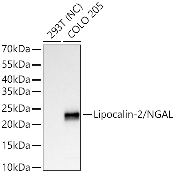
Western blot analysis of various lysates using Lipocalin-2/NGAL Rabbit PolymAb® at 1:1000dilution incubated overnight at 4℃.Secondary antibody: HRP-conjugated Goat anti-Rabbit IgG (H+L) at 1:10000 dilution.Lysates/proteins: 25 μg per lane.Blocking buffer: 3% nonfat dry milk in TBST.Detection: ECL Basic Kit.Negative control (NC): 293TExposure time: 10s.WB
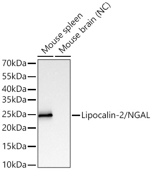
Western blot analysis of various lysates using Lipocalin-2/NGAL Rabbit PolymAb® at 1:1000dilution incubated overnight at 4℃.Secondary antibody: HRP-conjugated Goat anti-Rabbit IgG (H+L)at 1:10000 dilution.Lysates/proteins: 25 μg per lane.Blocking buffer: 3% nonfat dry milk in TBST.Detection: ECL Basic Kit.Negative control (NC): Mouse brainExposure time: 10s.WB
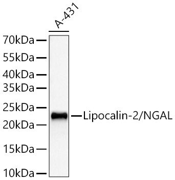
Western blot analysis of lysates from A-431 cells using Lipocalin-2/NGAL Rabbit PolymAb® at1:1000 dilution incubated overnight at 4℃.Secondary antibody: HRP-conjugated Goat anti-Rabbit IgG (H+L) at 1:10000 dilution.Lysates/proteins: 25 μg per lane.Blocking buffer: 3% nonfat dry milk in TBST.Detection: ECL Basic Kit .Exposure time: 60s.WB
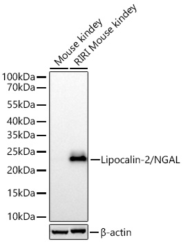
Western blot analysis of various lysates using Lipocalin-2/NGAL Rabbit PolymAb® at 1:1000dilution incubated overnight at 4℃.Secondary antibody: HRP-conjugated Goat anti-Rabbit IgG (H+L) at 1:10000 dilution.Lysates/proteins: 25 μg per lane.Blocking buffer: 3% nonfat dry milk in TBST.Detection: ECL Basic Kit.Exposure time: 20s.IHC
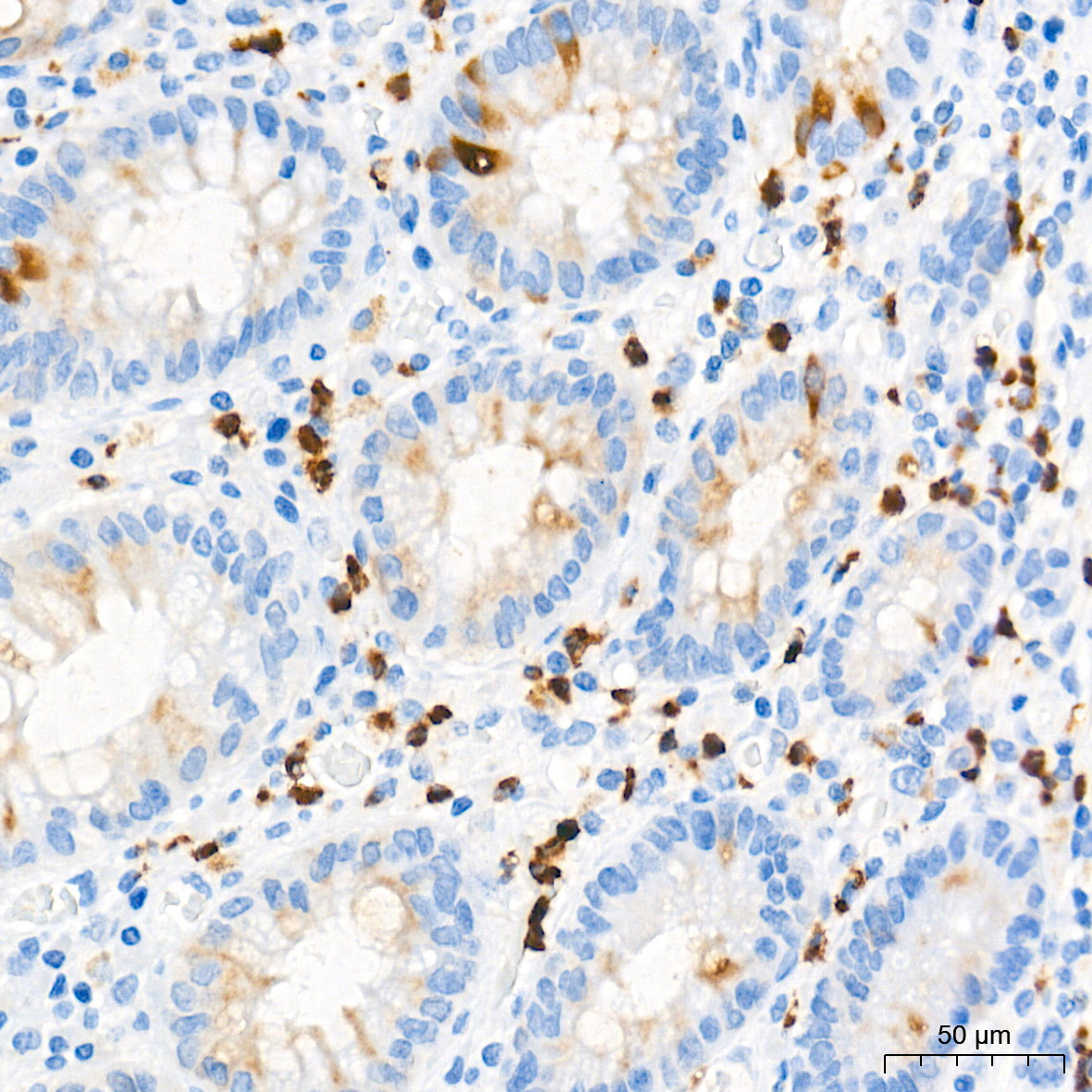
Immunohistochemistry analysis of paraffinembedded Human colon tissue usingLipocalin-2/NGAL Rabbit PolymAb® at a dilution of 1:1000 (40xlens). High pressure antigen retrievalperformed with 0.01M Tris-EDTA Buffer (pH9.0) prior to IHC staining.IHC
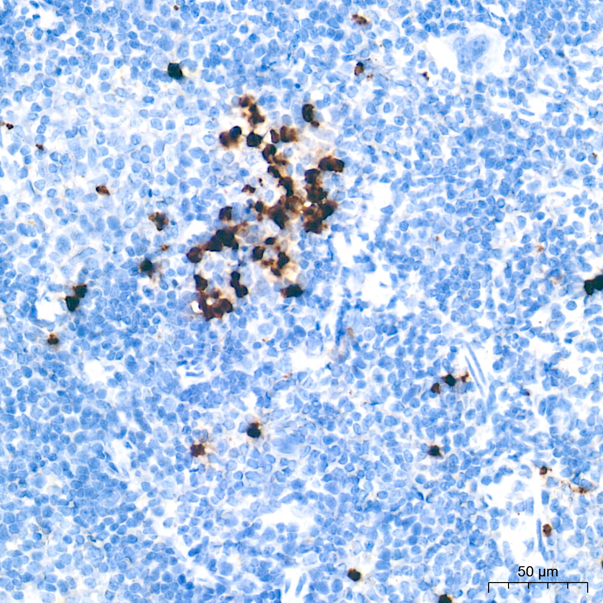
Immunohistochemistry analysis of paraffinembedded Mouse spleen tissue usingLipocalin-2/NGAL Rabbit PolymAb® at a dilution of 1:1000 (40xlens). High pressure antigen retrievalperformed with 0.01M Tris-EDTA Buffer (pH9.0) prior to IHC staining.IF-P
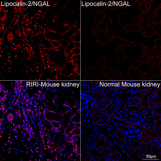
Confocal imaging of paraffin-embedded RIRIMouse kidney tissue (model for RenalIschemia-Reperfusion Injury, left) andC57BL6 Mouse kidney tissue (normal, right)using Lipocalin-2/NGAL Rabbit PolymAb®(dilution 1:200) followed by afurther incubation with Cy3-conjugated GoatAnti-Rabbit IgG (H+L) (dilution 1:500)(Red). DAPI was used for nuclear staining(Blue). High pressure antigen retrievalperformed with 0.01M Citrate Buffer (pH 6.0)prior to IF staining. Objective: 40x.IF-P
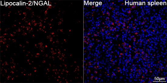
Confocal imaging of paraffin-embeddedHuman spleen tissue using Lipocalin-2/NGALRabbit PolymAb® (dilution1:200) followed by a further incubation withCy3-conjugated Goat Anti-Rabbit IgG (H+L)(dilution 1:500) (Red). DAPI was usedfor nuclear staining (Blue). High pressureantigen retrieval performed with 0.01MCitrate Buffer (pH 6.0) prior to IF staining.Objective: 40x.| Product Name | Lipocalin-2/NGAL Rabbit PolymAb |
|---|---|
| Antibody Type | Primary Antibodies |
| Immunogen | Recombinant protein (or fragment).This information is considered to be commercially sensitive. |
| Clonality | Polyclonal |
|---|---|
| Isotype | IgG |
| Host Species | Rabbit |
| Tested Applications | IF-PIHCWB |
| WB:1:1000-1:5000 IHC:1:500-1:2000 IF-P:1:200-1:400 |
|
| Species Reactivity | HumanMouse |
| Concentration | 1mg/ml |
| Purification | Affinity purified |
| Gene Symbol | LCN2 |
|---|---|
| Gene Synonyms | p25 24p3 MSFI NGAL |
| Gene Full Name | lipocalin 2 |
| Gene Summary | This gene encodes a protein that belongs to the lipocalin family. Members of this family transport small hydrophobic molecules such as lipids, steroid hormones and retinoids. The protein encoded by this gene is a neutrophil gelatinase-associated lipocalin and plays a role in innate immunity by limiting bacterial growth as a result of sequestering iron-containing siderophores. The presence of this protein in blood and urine is an early biomarker of acute kidney injury. This protein is thought to be be involved in multiple cellular processes, including maintenance of skin homeostasis, and suppression of invasiveness and metastasis. Mice lacking this gene are more susceptible to bacterial infection than wild type mice. [provided by RefSeq, Sep 2015] |
| Alternative Names | p25 24p3 MSFI NGAL Lipocalin-2/NGAL AW212229 NRL Sip24 |
| Molecular Weight(MW) | 23kDa(Observed band size:23kDa) |
| Cellular Localization | Secreted, Cytoplasmic granule lumen, Cytoplasmic vesicle lumen. |
WB
Western blot analysis of various lysates using Lipocalin-2/NGAL Rabbit PolymAb® at 1:1000dilution incubated overnight at 4℃.Secondary antibody: HRP-conjugated Goat anti-Rabbit IgG (H+L) at 1:10000 dilution.Lysates/proteins: 25 μg per lane.Blocking buffer: 3% nonfat dry milk in TBST.Detection: ECL Basic Kit.Negative control (NC): 293TExposure time: 10s.
WB
Western blot analysis of various lysates using Lipocalin-2/NGAL Rabbit PolymAb® at 1:1000dilution incubated overnight at 4℃.Secondary antibody: HRP-conjugated Goat anti-Rabbit IgG (H+L)at 1:10000 dilution.Lysates/proteins: 25 μg per lane.Blocking buffer: 3% nonfat dry milk in TBST.Detection: ECL Basic Kit.Negative control (NC): Mouse brainExposure time: 10s.
WB
Western blot analysis of lysates from A-431 cells using Lipocalin-2/NGAL Rabbit PolymAb® at1:1000 dilution incubated overnight at 4℃.Secondary antibody: HRP-conjugated Goat anti-Rabbit IgG (H+L) at 1:10000 dilution.Lysates/proteins: 25 μg per lane.Blocking buffer: 3% nonfat dry milk in TBST.Detection: ECL Basic Kit .Exposure time: 60s.
WB
Western blot analysis of various lysates using Lipocalin-2/NGAL Rabbit PolymAb® at 1:1000dilution incubated overnight at 4℃.Secondary antibody: HRP-conjugated Goat anti-Rabbit IgG (H+L) at 1:10000 dilution.Lysates/proteins: 25 μg per lane.Blocking buffer: 3% nonfat dry milk in TBST.Detection: ECL Basic Kit.Exposure time: 20s.
IHC
Immunohistochemistry analysis of paraffinembedded Human colon tissue usingLipocalin-2/NGAL Rabbit PolymAb® at a dilution of 1:1000 (40xlens). High pressure antigen retrievalperformed with 0.01M Tris-EDTA Buffer (pH9.0) prior to IHC staining.
IHC
Immunohistochemistry analysis of paraffinembedded Mouse spleen tissue usingLipocalin-2/NGAL Rabbit PolymAb® at a dilution of 1:1000 (40xlens). High pressure antigen retrievalperformed with 0.01M Tris-EDTA Buffer (pH9.0) prior to IHC staining.
IF-P
Confocal imaging of paraffin-embedded RIRIMouse kidney tissue (model for RenalIschemia-Reperfusion Injury, left) andC57BL6 Mouse kidney tissue (normal, right)using Lipocalin-2/NGAL Rabbit PolymAb®(dilution 1:200) followed by afurther incubation with Cy3-conjugated GoatAnti-Rabbit IgG (H+L) (dilution 1:500)(Red). DAPI was used for nuclear staining(Blue). High pressure antigen retrievalperformed with 0.01M Citrate Buffer (pH 6.0)prior to IF staining. Objective: 40x.
IF-P
Confocal imaging of paraffin-embeddedHuman spleen tissue using Lipocalin-2/NGALRabbit PolymAb® (dilution1:200) followed by a further incubation withCy3-conjugated Goat Anti-Rabbit IgG (H+L)(dilution 1:500) (Red). DAPI was usedfor nuclear staining (Blue). High pressureantigen retrieval performed with 0.01MCitrate Buffer (pH 6.0) prior to IF staining.Objective: 40x.| Application Notes | WB:1:1000-1:5000 IHC:1:500-1:2000 IF-P:1:200-1:400 |
|---|
| Form | Liquid |
|---|---|
| Storage Instructions | Store at -20℃. Avoid freeze / thaw cycles. |
| Storage Buffer | PBS with 0.09% Sodium azide, 0.05% BSA, 50% glycerol, pH7.3. |


