WB
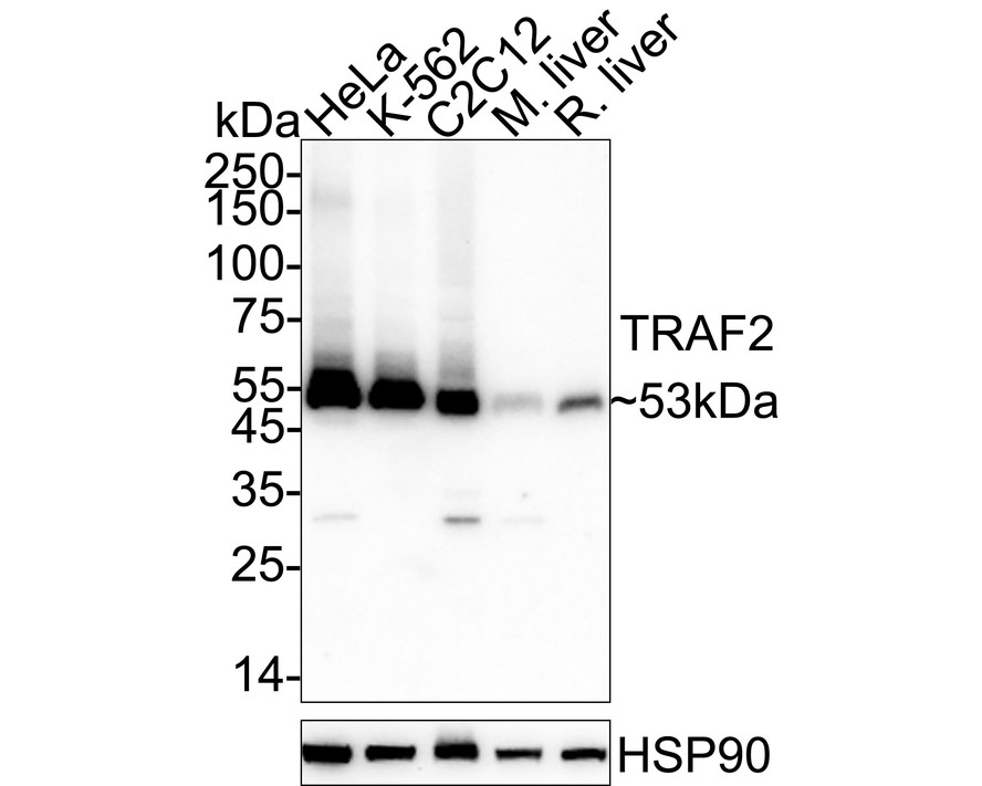
Western blot analysis of TRAF2 on different lysates with Rabbit anti-TRAF2 antibody at 1/1000 dilution. Lane 1: HeLa cell lysate (20 µg/Lane), Lane 2: K-562 cell lysate (20 µg/Lane), Lane 3: C2C12 cell lysate (20 µg/Lane), Lane 4: Mouse liver tissue lysate (40 µg/Lane), Lane 5: Rat liver tissue lysate (40 µg/Lane), Exposure time: 59 seconds;4-20% SDS-PAGE gel. Proteins were transferred to a PVDF membrane and blocked with 5% NFDM/TBST for 1 hour at room temperature. The primary antibody at 1/1000 dilution was used in 5% NFDM/TBST at 4℃ overnight. Goat Anti-Rabbit IgG - HRP Secondary Antibody at 1/50000 dilution was used for 1 hour at room temperature.IHC
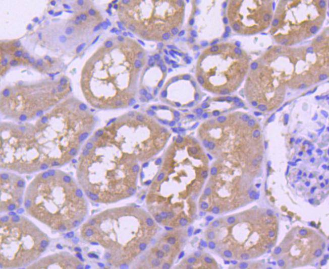
Immunohistochemical analysis of paraffin-embedded human kidney tissue using anti-TRAF2 antibody. The section was pre-treated using heat mediated antigen retrieval with Tris-EDTA buffer (pH 8.0-8.4) for 20 minutes.The tissues were blocked in 5% BSA for 30 minutes at room temperature, washed with ddH2O and PBS, and then probed with the primary antibody (1/50) for 30 minutes at room temperature. The detection was performed using an HRP conjugated compact polymer system. DAB was used as the chromogen. Tissues were counterstained with hematoxylin and mounted with DPX.IHC
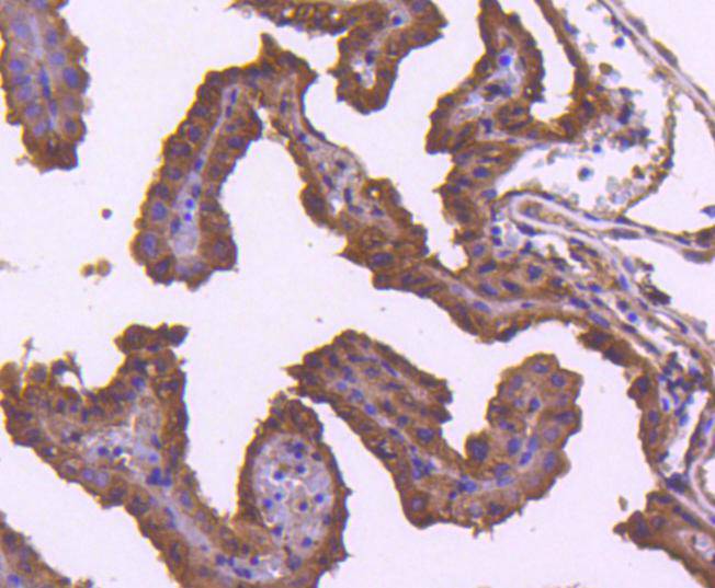
Immunohistochemical analysis of paraffin-embedded mouse placenta tissue using anti-TRAF2 antibody. The section was pre-treated using heat mediated antigen retrieval with Tris-EDTA buffer (pH 8.0-8.4) for 20 minutes.The tissues were blocked in 5% BSA for 30 minutes at room temperature, washed with ddH2O and PBS, and then probed with the primary antibody (1/50) for 30 minutes at room temperature. The detection was performed using an HRP conjugated compact polymer system. DAB was used as the chromogen. Tissues were counterstained with hematoxylin and mounted with DPX.| Product Name | TRAF2 Recombinant Rabbit Monoclonal Antibody |
|---|---|
| Antibody Type | Primary Antibodies |
| Immunogen | Synthetic peptide within N-terminal human TRAF2. |
| Clonality | Monoclonal |
|---|---|
| Isotype | IgG |
| Host Species | Rabbit |
| Tested Applications | IHCWB |
| WB:1:1000-1:5000 IHC:1:50-1:200 |
|
| Species Reactivity | HumanMouseRat |
| Concentration | 1mg/ml |
| Purification | Affinity purified |
| Gene Symbol | TRAF2 |
|---|---|
| Gene Synonyms | TRAP TRAP3 RNF117 MGC:45012 |
| Gene Full Name | TNF receptor associated factor 2 |
| Gene Summary | The protein encoded by this gene is a member of the TNF receptor associated factor (TRAF) protein family. TRAF proteins associate with, and mediate the signal transduction from members of the TNF receptor superfamily. This protein directly interacts with TNF receptors, and forms a heterodimeric complex with TRAF1. This protein is required for TNF-alpha-mediated activation of MAPK8/JNK and NF-kappaB. The protein complex formed by this protein and TRAF1 interacts with the inhibitor-of-apoptosis proteins (IAPs), and functions as a mediator of the anti-apoptotic signals from TNF receptors. The interaction of this protein with TRADD, a TNF receptor associated apoptotic signal transducer, ensures the recruitment of IAPs for the direct inhibition of caspase activation. BIRC2/c-IAP1, an apoptosis inhibitor possessing ubiquitin ligase activity, can unbiquitinate and induce the degradation of this protein, and thus potentiate TNF-induced apoptosis. Multiple alternatively spliced transcript variants have been found for this gene, but the biological validity of only one transcript has been determined. [provided by RefSeq, Jul 2008] |
| Alternative Names | E3 ubiquitin-protein ligase TRAF2 antibody MGC:45012 antibody OTTHUMP00000022625 antibody OTTHUMP00000064745 antibody TNF receptor associated factor 2 antibody TNF receptor-associated factor 2 antibody TNF receptor-associated protein antibody TRAF 2 antibody TRAF2 antibody TRAF2_HUMAN antibody E3 ubiquitin-protein ligase TRAF2 antibody MGC:45012 antibody OTTHUMP00000022625 antibody OTTHUMP00000064745 antibody TNF receptor associated factor 2 antibody TNF receptor-associated factor 2 antibody TNF receptor-associated protein antibody TRAF 2 antibody TRAF2 antibody TRAF2_HUMAN antibody TRAP 3 antibody TRAP antibody TRAP3 antibody Tumor necrosis factor type 2 receptor associated protein 3 antibody Tumor necrosis factor type 2 receptor-associated protein 3 antibody |
| Molecular Weight(MW) | 56kDa(Observed band size:53kDa) |
| Cellular Localization | Cytoplasm. |
WB
Western blot analysis of TRAF2 on different lysates with Rabbit anti-TRAF2 antibody at 1/1000 dilution. Lane 1: HeLa cell lysate (20 µg/Lane), Lane 2: K-562 cell lysate (20 µg/Lane), Lane 3: C2C12 cell lysate (20 µg/Lane), Lane 4: Mouse liver tissue lysate (40 µg/Lane), Lane 5: Rat liver tissue lysate (40 µg/Lane), Exposure time: 59 seconds;4-20% SDS-PAGE gel. Proteins were transferred to a PVDF membrane and blocked with 5% NFDM/TBST for 1 hour at room temperature. The primary antibody at 1/1000 dilution was used in 5% NFDM/TBST at 4℃ overnight. Goat Anti-Rabbit IgG - HRP Secondary Antibody at 1/50000 dilution was used for 1 hour at room temperature.
IHC
Immunohistochemical analysis of paraffin-embedded human kidney tissue using anti-TRAF2 antibody. The section was pre-treated using heat mediated antigen retrieval with Tris-EDTA buffer (pH 8.0-8.4) for 20 minutes.The tissues were blocked in 5% BSA for 30 minutes at room temperature, washed with ddH2O and PBS, and then probed with the primary antibody (1/50) for 30 minutes at room temperature. The detection was performed using an HRP conjugated compact polymer system. DAB was used as the chromogen. Tissues were counterstained with hematoxylin and mounted with DPX.
IHC
Immunohistochemical analysis of paraffin-embedded mouse placenta tissue using anti-TRAF2 antibody. The section was pre-treated using heat mediated antigen retrieval with Tris-EDTA buffer (pH 8.0-8.4) for 20 minutes.The tissues were blocked in 5% BSA for 30 minutes at room temperature, washed with ddH2O and PBS, and then probed with the primary antibody (1/50) for 30 minutes at room temperature. The detection was performed using an HRP conjugated compact polymer system. DAB was used as the chromogen. Tissues were counterstained with hematoxylin and mounted with DPX.| Application Notes | WB:1:1000-1:5000 IHC:1:50-1:200 |
|---|
| Form | Liquid |
|---|---|
| Storage Instructions | Shipped at 4℃. Store at +4℃ short term (1-2 weeks). It is recommended to aliquot into single-use upon delivery. Store at -20℃ long term. |
| Storage Buffer | 1*TBS (pH7.4), 0.05% BSA, 40% Glycerol. Preservative: 0.05% Sodium Azide. |


