WB
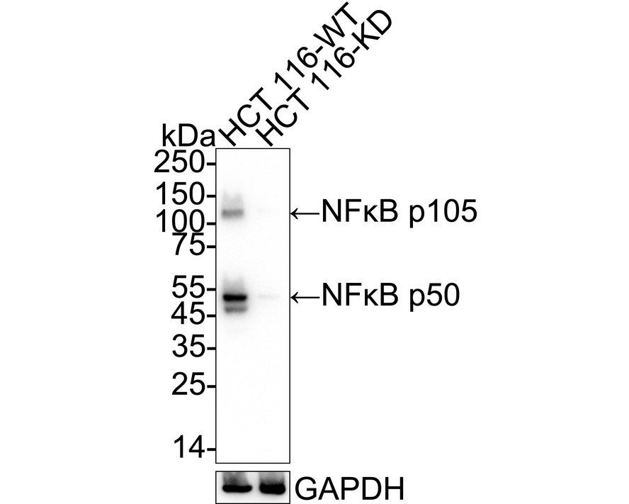
Western blot analysis of NF-kB p105 / p50 on different lysates with Rabbit anti-NF-kB p105 / p50 antibody at 1/5000 dilution. Lane 1: HCT 116-si NT cell lysate, Lane 2: HCT 116-si NFKB1 cell lysate, Lysates/proteins at 15 µg/Lane. Exposure time: 30 seconds; 4-20% SDS-PAGE gel. Proteins were transferred to a PVDF membrane and blocked with 5% NFDM/TBST for 1 hour at room temperature. The primary antibody at 1/5000 dilution was used in 5% NFDM/TBST at 4℃ overnight. Goat Anti-Rabbit IgG - HRP Secondary Antibody at 1/50000 dilution was used for 1 hour at room temperature.WB
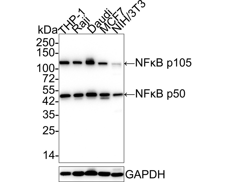
Western blot analysis of NF-kB p105 / p50 on different lysates with Rabbit anti-NF-kB p105 / p50 antibody at 1/5000 dilution. Lane 1: THP-1 cell lysate, Lane 2: Raji cell lysate, Lane 3: Daudi cell lysate, Lane 4: MCF7 cell lysate, Lane 5: NIH/3T3 cell lysate, Lysates/proteins at 15 µg/Lane. Exposure time: 3 minutes; 4-20% SDS-PAGE gel. Proteins were transferred to a PVDF membrane and blocked with 5% NFDM/TBST for 1 hour at room temperature. The primary antibody at 1/5000 dilution was used in 5% NFDM/TBST at 4℃ overnight. Goat Anti-Rabbit IgG - HRP Secondary Antibody at 1/50000 dilution was used for 1 hour at room temperature.WB
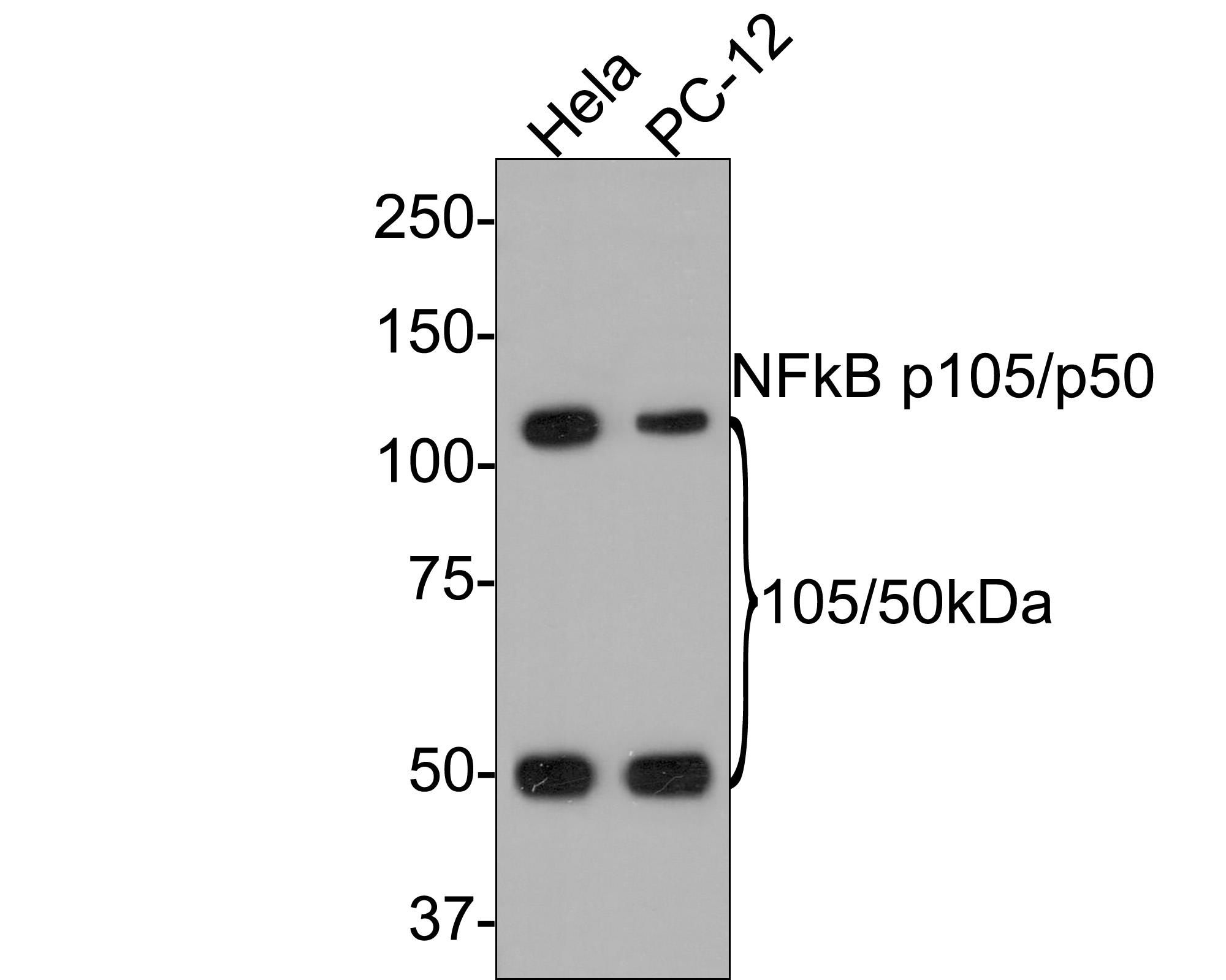
Western blot analysis of NF-kB p105 / p50 on different lysates with Rabbit anti-NF-kB p105 / p50 antibody at 1/500 dilution. Lane 1: Hela cell lysate, Lane 2: PC-12 cell lysate, Lysates/proteins at 10 µg/Lane. Exposure time: 2 minutes; 8% SDS-PAGE gel. Proteins were transferred to a PVDF membrane and blocked with 5% NFDM/TBST for 1 hour at room temperature. The primary antibody at 1/500 dilution was used in 5% NFDM/TBST at room temperature for 2 hours. Goat Anti-Rabbit IgG - HRP Secondary Antibody at 1:300000 dilution was used for 1 hour at room temperature.ICC/IF
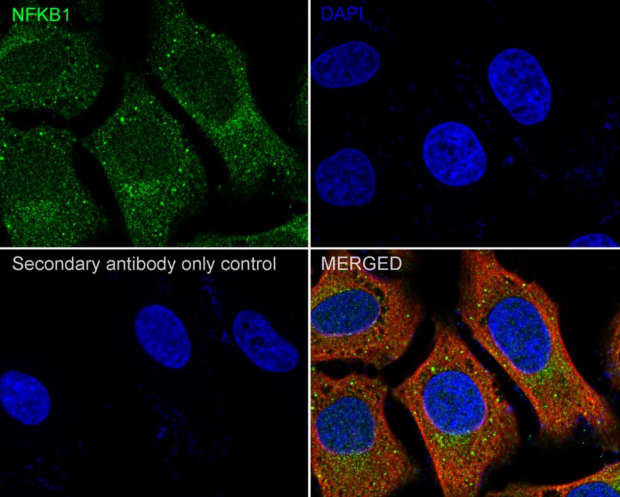
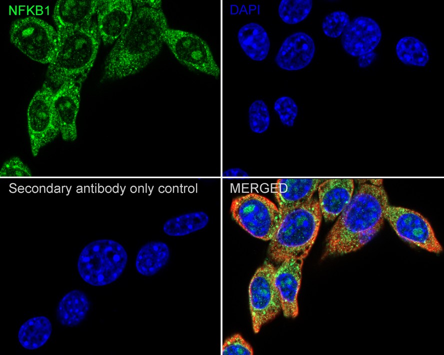
Immunocytochemistry analysis of HeLa cells labeling NF-kB p105 / p50 with Rabbit anti-NF-kB p105 / p50 antibody at 1/100 dilution. Cells were fixed in 4% paraformaldehyde for 20 minutes at room temperature, permeabilized with 0.1% Triton X-100 in PBS for 5 minutes at room temperature, then blocked with 1% BSA in 10% negative goat serum for 1 hour at room temperature. Cells were then incubated with Rabbit anti-NF-kB p105 / p50 antibody at 1/100 dilution in 1% BSA in PBST overnight at 4 ℃. Goat Anti-Rabbit IgG H&L (488) was used as the secondary antibody at 1/1,000 dilution. PBS instead of the primary antibody was used as the secondary antibody only control. Nuclear DNA was labelled in blue with DAPI. Beta tubulin (red) was stained at 1/100 dilution overnight at +4℃. Goat Anti-Mouse IgG H&L (594) was used as the secondary antibody at 1/1000 dilution.ICC/IF
Immunocytochemistry analysis of NIH/3T3 cells labeling NF-kB p105 / p50 with Rabbit anti-NF-kB p105 / p50 antibody at 1/100 dilution. Cells were fixed in 4% paraformaldehyde for 20 minutes at room temperature, permeabilized with 0.1% Triton X-100 in PBS for 5 minutes at room temperature, then blocked with 1% BSA in 10% negative goat serum for 1 hour at room temperature. Cells were then incubated with Rabbit anti-NF-kB p105 / p50 antibody at 1/100 dilution in 1% BSA in PBST overnight at 4 ℃. Goat Anti-Rabbit IgG H&L (488) was used as the secondary antibody at 1/1,000 dilution. PBS instead of the primary antibody was used as the secondary antibody only control. Nuclear DNA was labelled in blue with DAPI. Beta tubulin (red) was stained at 1/100 dilution overnight at +4℃. Goat Anti-Mouse IgG H&L ( 594) was used as the secondary antibody at 1/1000 dilution.IHC
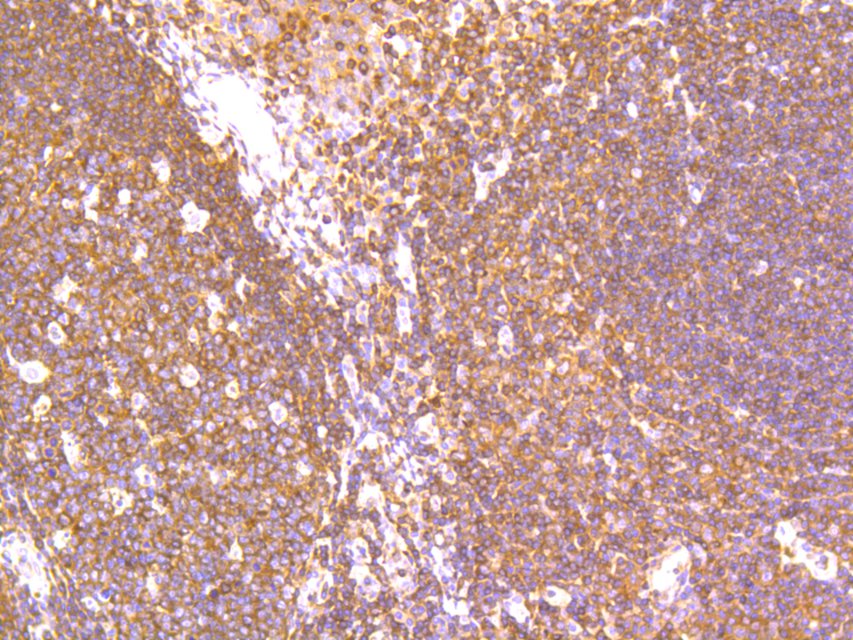
Immunohistochemical analysis of paraffin-embedded human tonsil tissue using anti-NF-kB p105 / p50 antibody. The section was pre-treated using heat mediated antigen retrieval with Tris-EDTA buffer (pH 8.0-8.4) for 20 minutes.The tissues were blocked in 5% BSA for 30 minutes at room temperature, washed with ddH2O and PBS, and then probed with the primary antibody (1/200) for 30 minutes at room temperature. The detection was performed using an HRP conjugated compact polymer system. DAB was used as the chromogen. Tissues were counterstained with hematoxylin and mounted with DPX.IHC
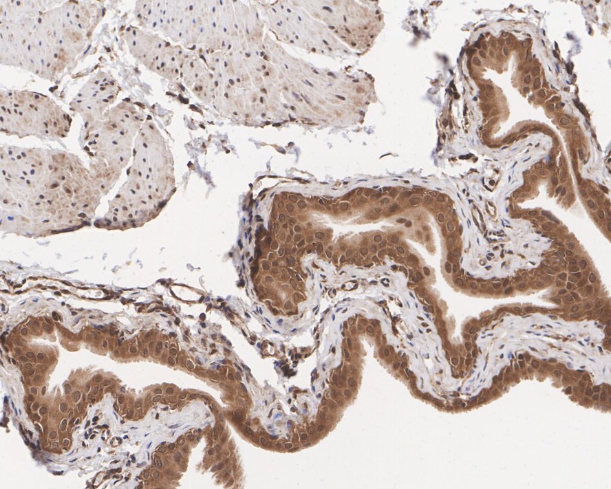
Immunohistochemical analysis of paraffin-embedded rat bladder tissue with Rabbit anti-NF-kB p105 / p50 antibody at 1/1000 dilution. The section was pre-treated using heat mediated antigen retrieval with Tris-EDTA buffer (pH 9.0) for 20 minutes. The tissues were blocked in 1% BSA for 20 minutes at room temperature, washed with ddH2O and PBS, and then probed with the primary antibody at 1/1000 dilution for 1 hour at room temperature. The detection was performed using an HRP conjugated compact polymer system. DAB was used as the chromogen. Tissues were counterstained with hematoxylin and mounted with DPX.FC
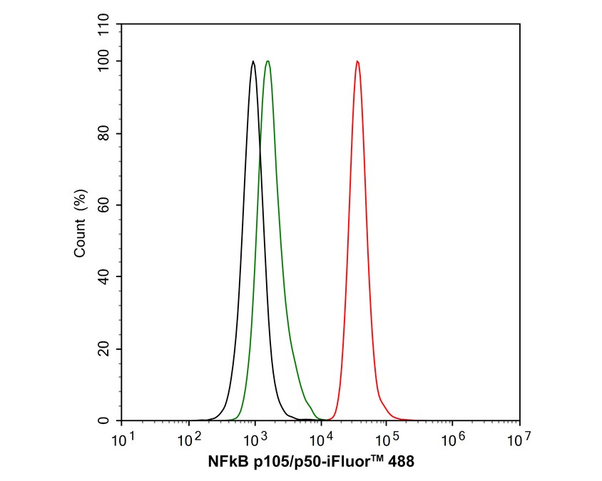
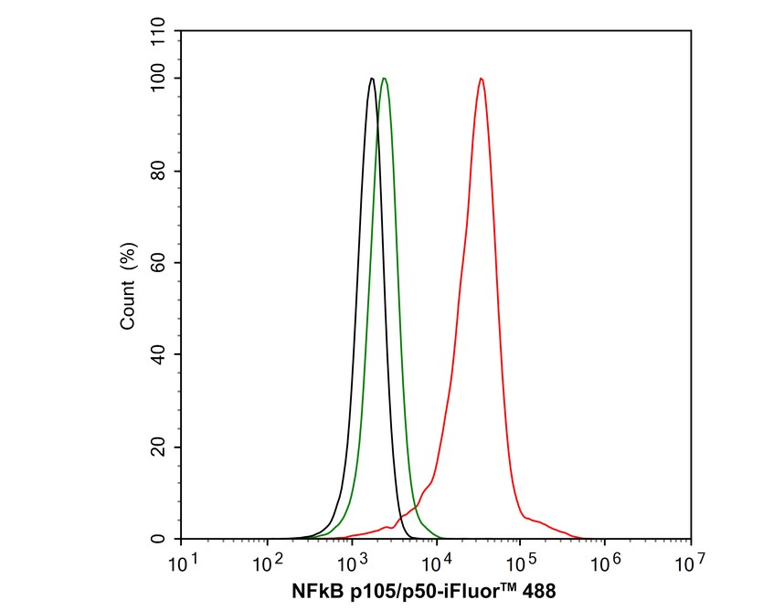
Flow cytometric analysis of HeLa cells labeling NF-kB p105 / p50. Cells were fixed and permeabilized. Then stained with the primary antibody (1μg/mL) (red) compared with Rabbit IgG Isotype Control (green). After incubation of the primary antibody at +4℃ for an hour, the cells were stained with a iFluor™ 488 conjugate-Goat anti-Rabbit IgG Secondary antibody at 1/1000 dilution for 30 minutes at +4℃. Unlabelled sample was used as a control (cells without incubation with primary antibody; black).FC
Flow cytometric analysis of NIH/3T3 cells labeling NF-kB p105 / p50. Cells were fixed and permeabilized. Then stained with the primary antibody (1μg/mL) (red) compared with Rabbit IgG Isotype Control (green). After incubation of the primary antibody at +4℃ for an hour, the cells were stained with a iFluor™ 488 conjugate-Goat anti-Rabbit IgG Secondary antibody at 1/1000 dilution for 30 minutes at +4℃. Unlabelled sample was used as a control (cells without incubation with primary antibody; black)."| Product Name | NF-kB p105 / p50 Recombinant Rabbit Monoclonal Antibody |
|---|---|
| Antibody Type | Primary Antibodies |
| Immunogen | Synthetic peptide within human NFKB1 aa 330-370. |
| Clonality | Monoclonal |
|---|---|
| Isotype | IgG |
| Host Species | Rabbit |
| Tested Applications | FCICC/IFIHCWB |
| WB:1:5000 IHC:1:200-1:1000 ICC/IF:1:100 FC:1:1000 |
|
| Species Reactivity | HumanMouseRat |
| Concentration | 1mg/ml |
| Purification | Affinity purified |
| Gene Symbol | NFKB1 |
|---|---|
| Gene Synonyms | KBF1 EBP-1 NF-kB CVID12 NF-kB1 NFKB-p50 NFkappaB NF-kappaB NFKB-p105 NF-kappa-B1 NF-kappabeta |
| Gene Full Name | nuclear factor kappa B subunit 1 |
| Gene Summary | This gene encodes a 105 kD protein which can undergo cotranslational processing by the 26S proteasome to produce a 50 kD protein. The 105 kD protein is a Rel protein-specific transcription inhibitor and the 50 kD protein is a DNA binding subunit of the NF-kappa-B (NFKB) protein complex. NFKB is a transcription regulator that is activated by various intra- and extra-cellular stimuli such as cytokines, oxidant-free radicals, ultraviolet irradiation, and bacterial or viral products. Activated NFKB translocates into the nucleus and stimulates the expression of genes involved in a wide variety of biological functions. Inappropriate activation of NFKB has been associated with a number of inflammatory diseases while persistent inhibition of NFKB leads to inappropriate immune cell development or delayed cell growth. NFKB is a critical regulator of the immediate-early response to viral infection. Alternative splicing results in multiple transcript variants encoding different isoforms, at least one of which is proteolytically processed. [provided by RefSeq, Aug 2020] |
| Alternative Names | DKFZp686C01211 antibody DNA binding factor KBF1 antibody DNA binding factor KBF1 EBP1 antibody DNA-binding factor KBF1 antibody EBP 1 antibody EBP-1 antibody EBP1 antibody KBF1 antibody MGC54151 antibody NF kappa B antibody DKFZp686C01211 antibody DNA binding factor KBF1 antibody DNA binding factor KBF1 EBP1 antibody DNA-binding factor KBF1 antibody EBP 1 antibody EBP-1 antibody EBP1 antibody KBF1 antibody MGC54151 antibody NF kappa B antibody NF kappaB antibody NF kappabeta antibody NF kB1 antibody NFkappaB antibody NFKB 1 antibody NFKB p105 antibody NFKB p50 antibody Nfkb1 antibody NFKB1_HUMAN antibody Nuclear factor kappa B DNA binding subunit antibody Nuclear factor kappa-B subunit 1 antibody Nuclear factor NF kappa B p105 subunit antibody Nuclear factor NF kappa B p50 subunit antibody Nuclear factor NF-kappa-B p50 subunit antibody Nuclear factor of kappa light chain gene enhancer in B cells 1 antibody Nuclear factor of kappa light polypeptide gene enhancer in B cells 1 antibody Nuclear factor of kappa light polypeptide gene enhancer in B-cells 1 antibody p105 antibody p50 antibody p84/NF-kappa-B1 p98 antibody Transcription factor NFKB1 antibody |
| Molecular Weight(MW) | 105/50kDa(Observed band size:105/50kDa) |
| Cellular Localization | Cytoplasm, Nucleus. |
WB
Western blot analysis of NF-kB p105 / p50 on different lysates with Rabbit anti-NF-kB p105 / p50 antibody at 1/5000 dilution. Lane 1: HCT 116-si NT cell lysate, Lane 2: HCT 116-si NFKB1 cell lysate, Lysates/proteins at 15 µg/Lane. Exposure time: 30 seconds; 4-20% SDS-PAGE gel. Proteins were transferred to a PVDF membrane and blocked with 5% NFDM/TBST for 1 hour at room temperature. The primary antibody at 1/5000 dilution was used in 5% NFDM/TBST at 4℃ overnight. Goat Anti-Rabbit IgG - HRP Secondary Antibody at 1/50000 dilution was used for 1 hour at room temperature.
WB
Western blot analysis of NF-kB p105 / p50 on different lysates with Rabbit anti-NF-kB p105 / p50 antibody at 1/5000 dilution. Lane 1: THP-1 cell lysate, Lane 2: Raji cell lysate, Lane 3: Daudi cell lysate, Lane 4: MCF7 cell lysate, Lane 5: NIH/3T3 cell lysate, Lysates/proteins at 15 µg/Lane. Exposure time: 3 minutes; 4-20% SDS-PAGE gel. Proteins were transferred to a PVDF membrane and blocked with 5% NFDM/TBST for 1 hour at room temperature. The primary antibody at 1/5000 dilution was used in 5% NFDM/TBST at 4℃ overnight. Goat Anti-Rabbit IgG - HRP Secondary Antibody at 1/50000 dilution was used for 1 hour at room temperature.
WB
Western blot analysis of NF-kB p105 / p50 on different lysates with Rabbit anti-NF-kB p105 / p50 antibody at 1/500 dilution. Lane 1: Hela cell lysate, Lane 2: PC-12 cell lysate, Lysates/proteins at 10 µg/Lane. Exposure time: 2 minutes; 8% SDS-PAGE gel. Proteins were transferred to a PVDF membrane and blocked with 5% NFDM/TBST for 1 hour at room temperature. The primary antibody at 1/500 dilution was used in 5% NFDM/TBST at room temperature for 2 hours. Goat Anti-Rabbit IgG - HRP Secondary Antibody at 1:300000 dilution was used for 1 hour at room temperature.
ICC/IF
Immunocytochemistry analysis of HeLa cells labeling NF-kB p105 / p50 with Rabbit anti-NF-kB p105 / p50 antibody at 1/100 dilution. Cells were fixed in 4% paraformaldehyde for 20 minutes at room temperature, permeabilized with 0.1% Triton X-100 in PBS for 5 minutes at room temperature, then blocked with 1% BSA in 10% negative goat serum for 1 hour at room temperature. Cells were then incubated with Rabbit anti-NF-kB p105 / p50 antibody at 1/100 dilution in 1% BSA in PBST overnight at 4 ℃. Goat Anti-Rabbit IgG H&L (488) was used as the secondary antibody at 1/1,000 dilution. PBS instead of the primary antibody was used as the secondary antibody only control. Nuclear DNA was labelled in blue with DAPI. Beta tubulin (red) was stained at 1/100 dilution overnight at +4℃. Goat Anti-Mouse IgG H&L (594) was used as the secondary antibody at 1/1000 dilution.
ICC/IF
Immunocytochemistry analysis of NIH/3T3 cells labeling NF-kB p105 / p50 with Rabbit anti-NF-kB p105 / p50 antibody at 1/100 dilution. Cells were fixed in 4% paraformaldehyde for 20 minutes at room temperature, permeabilized with 0.1% Triton X-100 in PBS for 5 minutes at room temperature, then blocked with 1% BSA in 10% negative goat serum for 1 hour at room temperature. Cells were then incubated with Rabbit anti-NF-kB p105 / p50 antibody at 1/100 dilution in 1% BSA in PBST overnight at 4 ℃. Goat Anti-Rabbit IgG H&L (488) was used as the secondary antibody at 1/1,000 dilution. PBS instead of the primary antibody was used as the secondary antibody only control. Nuclear DNA was labelled in blue with DAPI. Beta tubulin (red) was stained at 1/100 dilution overnight at +4℃. Goat Anti-Mouse IgG H&L ( 594) was used as the secondary antibody at 1/1000 dilution.
IHC
Immunohistochemical analysis of paraffin-embedded human tonsil tissue using anti-NF-kB p105 / p50 antibody. The section was pre-treated using heat mediated antigen retrieval with Tris-EDTA buffer (pH 8.0-8.4) for 20 minutes.The tissues were blocked in 5% BSA for 30 minutes at room temperature, washed with ddH2O and PBS, and then probed with the primary antibody (1/200) for 30 minutes at room temperature. The detection was performed using an HRP conjugated compact polymer system. DAB was used as the chromogen. Tissues were counterstained with hematoxylin and mounted with DPX.
IHC
Immunohistochemical analysis of paraffin-embedded rat bladder tissue with Rabbit anti-NF-kB p105 / p50 antibody at 1/1000 dilution. The section was pre-treated using heat mediated antigen retrieval with Tris-EDTA buffer (pH 9.0) for 20 minutes. The tissues were blocked in 1% BSA for 20 minutes at room temperature, washed with ddH2O and PBS, and then probed with the primary antibody at 1/1000 dilution for 1 hour at room temperature. The detection was performed using an HRP conjugated compact polymer system. DAB was used as the chromogen. Tissues were counterstained with hematoxylin and mounted with DPX.
FC
Flow cytometric analysis of HeLa cells labeling NF-kB p105 / p50. Cells were fixed and permeabilized. Then stained with the primary antibody (1μg/mL) (red) compared with Rabbit IgG Isotype Control (green). After incubation of the primary antibody at +4℃ for an hour, the cells were stained with a iFluor™ 488 conjugate-Goat anti-Rabbit IgG Secondary antibody at 1/1000 dilution for 30 minutes at +4℃. Unlabelled sample was used as a control (cells without incubation with primary antibody; black).
FC
Flow cytometric analysis of NIH/3T3 cells labeling NF-kB p105 / p50. Cells were fixed and permeabilized. Then stained with the primary antibody (1μg/mL) (red) compared with Rabbit IgG Isotype Control (green). After incubation of the primary antibody at +4℃ for an hour, the cells were stained with a iFluor™ 488 conjugate-Goat anti-Rabbit IgG Secondary antibody at 1/1000 dilution for 30 minutes at +4℃. Unlabelled sample was used as a control (cells without incubation with primary antibody; black)."| Application Notes | WB:1:5000 IHC:1:200-1:1000 ICC/IF:1:100 FC:1:1000 |
|---|
| Form | Liquid |
|---|---|
| Storage Instructions | Shipped at 4℃. Store at +4℃ short term (1-2 weeks). It is recommended to aliquot into single-use upon delivery. Store at -20℃ long term. |
| Storage Buffer | 1*TBS (pH7.4), 0.05% BSA, 40% Glycerol. Preservative: 0.05% Sodium Azide. |


