WB
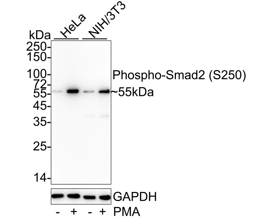
Western blot analysis of Phospho-Smad2 (S250) on different lysates with Rabbit anti-Phospho-Smad2 (S250) antibody at 1/5000 dilution. Lane 1: HeLa cell lysate, Lane 2: HeLa treated with 200nM PMA for 30 minutes cell lysate, Lane 3: NIH/3T3 cell lysate, Lane 4: NIH/3T3 treated with 200nM PMA for 30 minutes cell lysate, Lysates/proteins at 20 µg/Lane. Exposure time: 30 seconds; 4-20% SDS-PAGE gel. Proteins were transferred to a PVDF membrane and blocked with 5% NFDM/TBST for 1 hour at room temperature. The primary antibody at 1/5000 dilution was used in 5% NFDM/TBST at 4℃ overnight. Goat Anti-Rabbit IgG - HRP Secondary Antibody at 1/50000 dilution was used for 1 hour at room temperature.WB
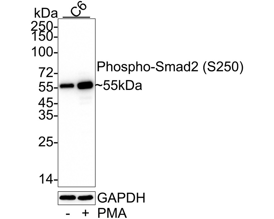
Western blot analysis of Phospho-Smad2 (S250) on different lysates with Rabbit anti-Phospho-Smad2 (S250) antibody at 1/5000 dilution. Lane 1: C6 cell lysate, Lane 2: C6 treated with 200nM PMA for 30 minutes cell lysate, Lysates/proteins at 20 µg/Lane. Exposure time: 2 minutes; 4-20% SDS-PAGE gel. Proteins were transferred to a PVDF membrane and blocked with 5% NFDM/TBST for 1 hour at room temperature. The primary antibody at 1/5000 dilution was used in 5% NFDM/TBST at 4℃ overnight. Goat Anti-Rabbit IgG - HRP Secondary Antibody at 1/50000 dilution was used for 1 hour at room temperature.IP
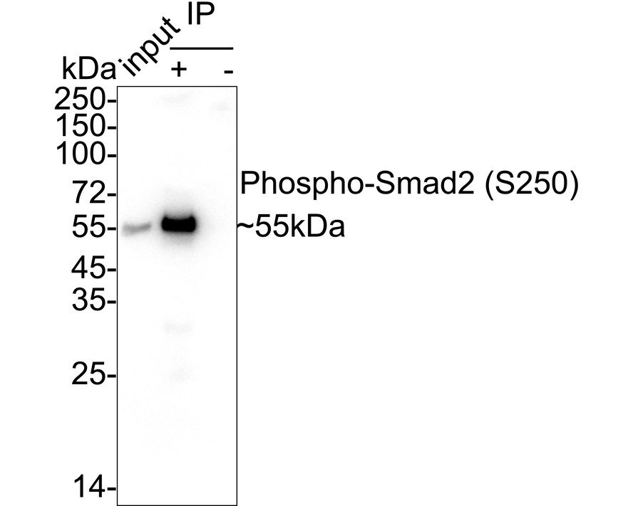
Phospho-Smad2 (S250) was immunoprecipitated from 0.2 mg HeLa treated with 100nM TPA for 30 minutes cell lysate with Rabbit anti-Phospho-Smad2 (S250) antibody at 2 µg/10 µl beads. Western blot was performed from the immunoprecipitate using Rabbit anti-Phospho-Smad2 (S250) antibody at 1/5000 dilution. Anti-Rabbit IgG for IP Nano-secondary antibody at 1/5000 dilution was used for 1 hour at room temperature. Lane 1: HeLa treated with 100nM TPA for 30 minutes cell lysate (input), Lane 2: Rabbit anti-Phospho-Smad2 (S250) antibody IP in HeLa treated with 100nM TPA for 30 minutes cell lysate, Lane 3: Rabbit IgG instead of Rabbit anti-Phospho-Smad2 (S250) antibody in HeLa treated with 100nM TPA for 30 minutes cell lysate, Blocking/Dilution buffer: 5% NFDM/TBST, Exposure time: 35 seconds.IHC
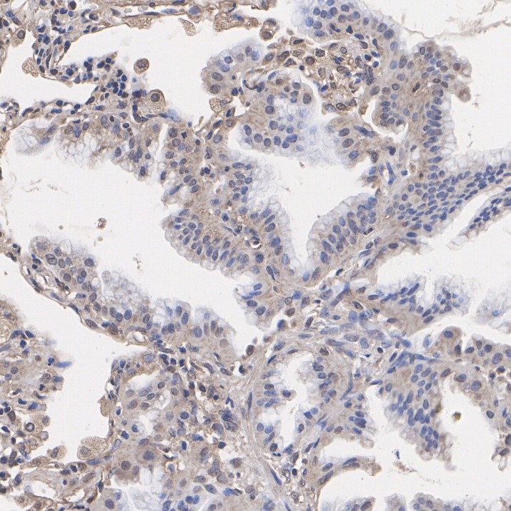
Immunohistochemical analysis of paraffin-embedded human lung cancer tissue with Rabbit anti-Phospho-Smad2 (S250) antibody at 1/50 dilution. The section was pre-treated using heat mediated antigen retrieval with Tris-EDTA buffer (pH 9.0) for 20 minutes. The tissues were blocked in 1% BSA for 20 minutes at room temperature, washed with ddH2O and PBS, and then probed with the primary antibody at 1/50 dilution for 1 hour at room temperature. The detection was performed using an HRP conjugated compact polymer system. DAB was used as the chromogen. Tissues were counterstained with hematoxylin and mounted with DPX.IHC
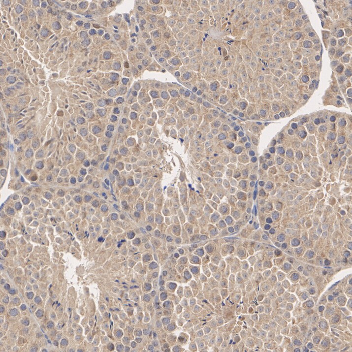
Immunohistochemical analysis of paraffin-embedded mouse testis tissue with Rabbit anti-Phospho-Smad2 (S250) antibody at 1/50 dilution. The section was pre-treated using heat mediated antigen retrieval with Tris-EDTA buffer (pH 9.0) for 20 minutes. The tissues were blocked in 1% BSA for 20 minutes at room temperature, washed with ddH2O and PBS, and then probed with the primary antibody at 1/50 dilution for 1 hour at room temperature. The detection was performed using an HRP conjugated compact polymer system. DAB was used as the chromogen. Tissues were counterstained with hematoxylin and mounted with DPX.IHC
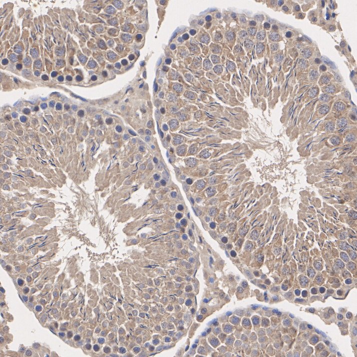
Immunohistochemical analysis of paraffin-embedded rat testis tissue with Rabbit anti-Phospho-Smad2 (S250) antibody at 1/50 dilution. The section was pre-treated using heat mediated antigen retrieval with Tris-EDTA buffer (pH 9.0) for 20 minutes. The tissues were blocked in 1% BSA for 20 minutes at room temperature, washed with ddH2O and PBS, and then probed with the primary antibody at 1/50 dilution for 1 hour at room temperature. The detection was performed using an HRP conjugated compact polymer system. DAB was used as the chromogen. Tissues were counterstained with hematoxylin and mounted with DPX.FC
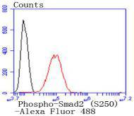
Flow cytometric analysis of HeLa cells labeling Phospho-Smad2 (S250). Cells were fixed and permeabilized. Then stained with the primary antibody (1/1000) (red) compared with Rabbit IgG Isotype Control (green). After incubation of the primary antibody at +4℃ for an hour, the cells were stained with a iFluor™ 488 conjugate-Goat anti-Rabbit IgG Secondary antibody at 1/1000 dilution for 30 minutes at +4℃. Unlabelled sample was used as a control (cells without incubation with primary antibody; black).| Product Name | Phospho-Smad2 (S250) Recombinant Rabbit Monoclonal Antibody |
|---|---|
| Antibody Type | Primary Antibodies |
| Immunogen | Synthetic phospho-peptide corresponding to residues surrounding Ser250 of Human Smad2 aa 221-270 / 467. |
| Modification | P-S250 |
| Clonality | Monoclonal |
|---|---|
| Isotype | IgG |
| Host Species | Rabbit |
| Tested Applications | FCIHCIPWB |
| WB:1:5000 IP:1-2μg/sample IHC:1:50 FC:1:50-1:100 |
|
| Species Reactivity | HumanMouseRat |
| Concentration | 1mg/ml |
| Purification | Affinity purified |
| Gene Symbol | SMAD2 |
|---|---|
| Gene Synonyms | JV18 LDS6 CHTD8 MADH2 MADR2 JV18-1 hMAD-2 hSMAD2 |
| Gene Full Name | SMAD family member 2 |
| Gene Summary | The protein encoded by this gene belongs to the SMAD, a family of proteins similar to the gene products of the Drosophila gene 'mothers against decapentaplegic' (Mad) and the C. elegans gene Sma. SMAD proteins are signal transducers and transcriptional modulators that mediate multiple signaling pathways. This protein mediates the signal of the transforming growth factor (TGF)-beta, and thus regulates multiple cellular processes, such as cell proliferation, apoptosis, and differentiation. This protein is recruited to the TGF-beta receptors through its interaction with the SMAD anchor for receptor activation (SARA) protein. In response to TGF-beta signal, this protein is phosphorylated by the TGF-beta receptors. The phosphorylation induces the dissociation of this protein with SARA and the association with the family member SMAD4. The association with SMAD4 is important for the translocation of this protein into the nucleus, where it binds to target promoters and forms a transcription repressor complex with other cofactors. This protein can also be phosphorylated by activin type 1 receptor kinase, and mediates the signal from the activin. Alternatively spliced transcript variants have been observed for this gene. [provided by RefSeq, May 2012] |
| Alternative Names | Drosophila homolog of MADR2 antibody hMAD-2 antibody HsMAD2 antibody JV18 antibody JV18-1 antibody JV181 antibody MAD antibody MAD homolog 2 antibody MAD Related Protein 2 antibody Mad-related protein 2 antibody Drosophila homolog of MADR2 antibody hMAD-2 antibody HsMAD2 antibody JV18 antibody JV18-1 antibody JV181 antibody MAD antibody MAD homolog 2 antibody MAD Related Protein 2 antibody Mad-related protein 2 antibody MADH2 antibody MADR2 antibody MGC22139 antibody MGC34440 antibody Mother against DPP homolog 2 antibody Mothers against decapentaplegic homolog 2 antibody Mothers against decapentaplegic Drosophila homolog of 2 antibody Mothers against DPP homolog 2 antibody OTTHUMP00000163489 antibody Sma and Mad related protein 2 antibody Sma- and Mad-related protein 2 MAD antibody SMAD 2 antibody SMAD family member 2 antibody SMAD mothers against DPP homolog 2 antibody SMAD2 antibody SMAD2_HUMAN antibody |
| Molecular Weight(MW) | 52kDa(Observed band size:55kDa) |
| Cellular Localization | Cytoplasm, Nucleus. |
WB
Western blot analysis of Phospho-Smad2 (S250) on different lysates with Rabbit anti-Phospho-Smad2 (S250) antibody at 1/5000 dilution. Lane 1: HeLa cell lysate, Lane 2: HeLa treated with 200nM PMA for 30 minutes cell lysate, Lane 3: NIH/3T3 cell lysate, Lane 4: NIH/3T3 treated with 200nM PMA for 30 minutes cell lysate, Lysates/proteins at 20 µg/Lane. Exposure time: 30 seconds; 4-20% SDS-PAGE gel. Proteins were transferred to a PVDF membrane and blocked with 5% NFDM/TBST for 1 hour at room temperature. The primary antibody at 1/5000 dilution was used in 5% NFDM/TBST at 4℃ overnight. Goat Anti-Rabbit IgG - HRP Secondary Antibody at 1/50000 dilution was used for 1 hour at room temperature.
WB
Western blot analysis of Phospho-Smad2 (S250) on different lysates with Rabbit anti-Phospho-Smad2 (S250) antibody at 1/5000 dilution. Lane 1: C6 cell lysate, Lane 2: C6 treated with 200nM PMA for 30 minutes cell lysate, Lysates/proteins at 20 µg/Lane. Exposure time: 2 minutes; 4-20% SDS-PAGE gel. Proteins were transferred to a PVDF membrane and blocked with 5% NFDM/TBST for 1 hour at room temperature. The primary antibody at 1/5000 dilution was used in 5% NFDM/TBST at 4℃ overnight. Goat Anti-Rabbit IgG - HRP Secondary Antibody at 1/50000 dilution was used for 1 hour at room temperature.
IP
Phospho-Smad2 (S250) was immunoprecipitated from 0.2 mg HeLa treated with 100nM TPA for 30 minutes cell lysate with Rabbit anti-Phospho-Smad2 (S250) antibody at 2 µg/10 µl beads. Western blot was performed from the immunoprecipitate using Rabbit anti-Phospho-Smad2 (S250) antibody at 1/5000 dilution. Anti-Rabbit IgG for IP Nano-secondary antibody at 1/5000 dilution was used for 1 hour at room temperature. Lane 1: HeLa treated with 100nM TPA for 30 minutes cell lysate (input), Lane 2: Rabbit anti-Phospho-Smad2 (S250) antibody IP in HeLa treated with 100nM TPA for 30 minutes cell lysate, Lane 3: Rabbit IgG instead of Rabbit anti-Phospho-Smad2 (S250) antibody in HeLa treated with 100nM TPA for 30 minutes cell lysate, Blocking/Dilution buffer: 5% NFDM/TBST, Exposure time: 35 seconds.
IHC
Immunohistochemical analysis of paraffin-embedded human lung cancer tissue with Rabbit anti-Phospho-Smad2 (S250) antibody at 1/50 dilution. The section was pre-treated using heat mediated antigen retrieval with Tris-EDTA buffer (pH 9.0) for 20 minutes. The tissues were blocked in 1% BSA for 20 minutes at room temperature, washed with ddH2O and PBS, and then probed with the primary antibody at 1/50 dilution for 1 hour at room temperature. The detection was performed using an HRP conjugated compact polymer system. DAB was used as the chromogen. Tissues were counterstained with hematoxylin and mounted with DPX.
IHC
Immunohistochemical analysis of paraffin-embedded mouse testis tissue with Rabbit anti-Phospho-Smad2 (S250) antibody at 1/50 dilution. The section was pre-treated using heat mediated antigen retrieval with Tris-EDTA buffer (pH 9.0) for 20 minutes. The tissues were blocked in 1% BSA for 20 minutes at room temperature, washed with ddH2O and PBS, and then probed with the primary antibody at 1/50 dilution for 1 hour at room temperature. The detection was performed using an HRP conjugated compact polymer system. DAB was used as the chromogen. Tissues were counterstained with hematoxylin and mounted with DPX.
IHC
Immunohistochemical analysis of paraffin-embedded rat testis tissue with Rabbit anti-Phospho-Smad2 (S250) antibody at 1/50 dilution. The section was pre-treated using heat mediated antigen retrieval with Tris-EDTA buffer (pH 9.0) for 20 minutes. The tissues were blocked in 1% BSA for 20 minutes at room temperature, washed with ddH2O and PBS, and then probed with the primary antibody at 1/50 dilution for 1 hour at room temperature. The detection was performed using an HRP conjugated compact polymer system. DAB was used as the chromogen. Tissues were counterstained with hematoxylin and mounted with DPX.
FC
Flow cytometric analysis of HeLa cells labeling Phospho-Smad2 (S250). Cells were fixed and permeabilized. Then stained with the primary antibody (1/1000) (red) compared with Rabbit IgG Isotype Control (green). After incubation of the primary antibody at +4℃ for an hour, the cells were stained with a iFluor™ 488 conjugate-Goat anti-Rabbit IgG Secondary antibody at 1/1000 dilution for 30 minutes at +4℃. Unlabelled sample was used as a control (cells without incubation with primary antibody; black).| Application Notes | WB:1:5000 IP:1-2μg/sample IHC:1:50 FC:1:50-1:100 |
|---|
| Form | Liquid |
|---|---|
| Storage Instructions | Shipped at 4℃. Store at +4℃ short term (1-2 weeks). It is recommended to aliquot into single-use upon delivery. Store at -20℃ long term. |
| Storage Buffer | 1*TBS (pH7.4), 0.05% BSA, 40% Glycerol. Preservative: 0.05% Sodium Azide. |


