WB
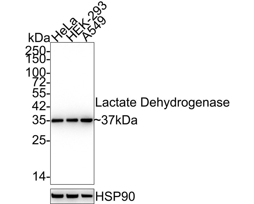
Western blot analysis of Lactate Dehydrogenase on different lysates with Rabbit anti-Lactate Dehydrogenase antibody at 1/5000 dilution. Lane 1: HeLa cell lysate (15 µg/Lane), Lane 2: HEK-293 cell lysate (15 µg/Lane), Lane 3: A549 cell lysate (15 µg/Lane), Exposure time: 1 minute 36 seconds; 4-20% SDS-PAGE gel. Proteins were transferred to a PVDF membrane and blocked with 5% NFDM/TBST for 1 hour at room temperature. The primary antibody at 1/5000 dilution was used in 5% NFDM/TBST at 4℃ overnight. Goat Anti-Rabbit IgG - HRP Secondary Antibody at 1:50000 dilution was used for 1 hour at room temperature.WB
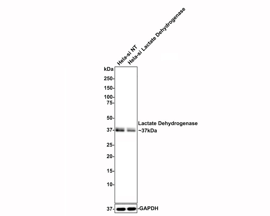
Western blot analysis of Lactate Dehydrogenase on different lysates with Rabbit anti-Lactate Dehydrogenase antibody at 1/1000 dilution. Lane 1: Hela-si NT cell lysate (10 µg/Lane), Lane 2: Hela-si Lactate Dehydrogenase cell lysate (10 µg/Lane), Exposure time: 25 seconds; 4-20% SDS-PAGE gel. Rabbit anti-Lactate Dehydrogenase antibody was shown to specifically react with Lactate Dehydrogenase in Hela-si NT cells. Weakened band was observed when Hela-si Lactate Dehydrogenase sample was tested. Hela-si NT and Hela-si Lactate Dehydrogenase samples were subjected to SDS-PAGE. Proteins were transferred to a PVDF membrane and blocked with 5% NFDM in TBST for 1 hour at room temperature. The primary antibody (1/1000) and Loading control antibody (Rabbit anti-GAPDH, 1/10000) were used in 5% BSA at room temperature for 2 hours. Goat Anti-rabbit IgG-HRP Secondary Antibody at 1:100000 dilution was used for 1 hour at room temperature.WB
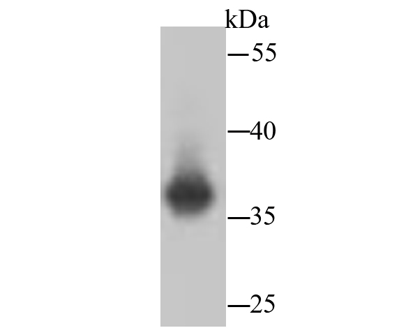
Western blot analysis of Lactate Dehydrogenase on hybrid fish (crucian-carp) brain tissue lysates. Proteins were transferred to a PVDF membrane and blocked with 5% BSA in PBS for 1 hour at room temperature. The primary antibody (1/500) was used in 5% BSA at room temperature for 2 hours. Goat Anti-Rabbit IgG - HRP Secondary Antibody at 1:200000 dilution was used for 1 hour at room temperature.ICC/IF
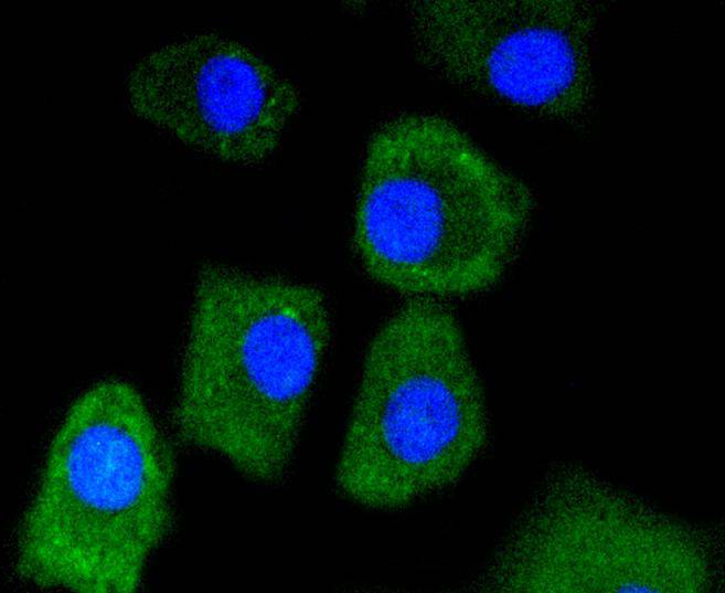
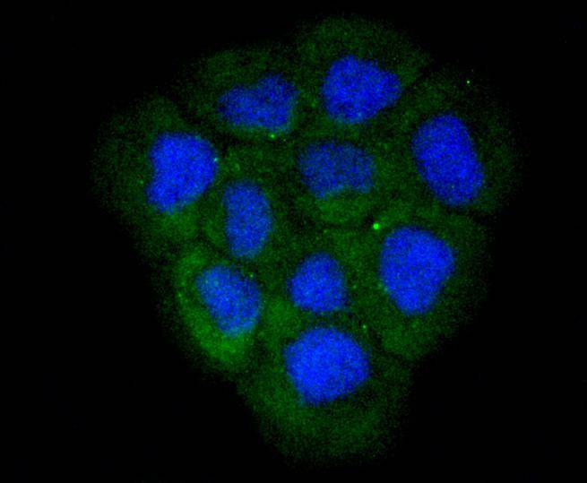
ICC staining of Lactate Dehydrogenase in A549 cells (green). Formalin fixed cells were permeabilized with 0.1% Triton X-100 in TBS for 10 minutes at room temperature and blocked with 10% negative goat serum for 15 minutes at room temperature. Cells were probed with the primary antibody (1/50) for 1 hour at room temperature, washed with PBS. Alexa Fluor®488 conjugate-Goat anti-Rabbit IgG was used as the secondary antibody at 1/1000 dilution. The nuclear counter stain is DAPI (blue).ICC/IF
ICC staining of Lactate Dehydrogenase in A431 cells (green). Formalin fixed cells were permeabilized with 0.1% Triton X-100 in TBS for 10 minutes at room temperature and blocked with 10% negative goat serum for 15 minutes at room temperature. Cells were probed with the primary antibody (1/50) for 1 hour at room temperature, washed with PBS. Alexa Fluor®488 conjugate-Goat anti-Rabbit IgG was used as the secondary antibody at 1/1000 dilution. The nuclear counter stain is DAPI (blue).ICC/IF
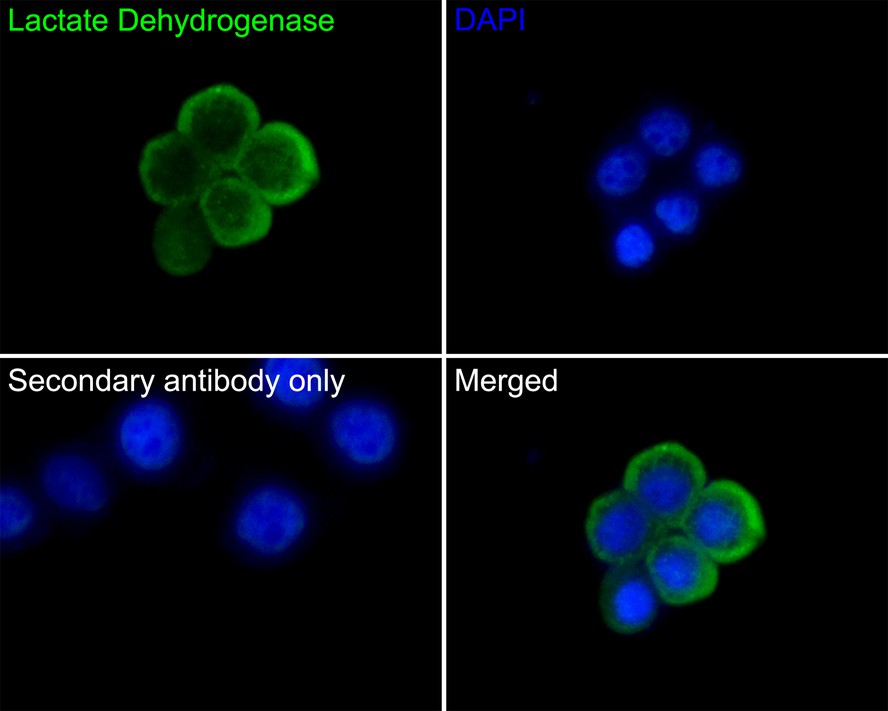
Immunocytochemistry analysis of LOVO cells labeling Lactate Dehydrogenase with Rabbit anti-Lactate Dehydrogenase antibody at 1/50 dilution. Cells were fixed in 4% paraformaldehyde for 10 minutes at 37 ℃, permeabilized with 0.05% Triton X-100 in PBS for 20 minutes, and then blocked with 2% negative goat serum for 30 minutes at room temperature. Cells were then incubated with Rabbit anti-Lactate Dehydrogenase antibody at 1/50 dilution in 2% negative goat serum overnight at 4 ℃. Goat Anti-Rabbit IgG H&L (488) was used as the secondary antibody at 1/1,000 dilution. PBS instead of the primary antibody was used as the secondary antibody only control. Nuclear DNA was labelled in blue with DAPI.IHC
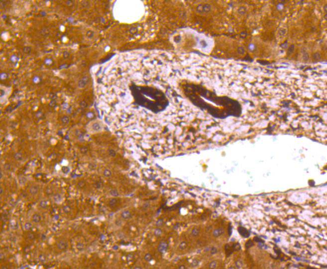
Immunohistochemical analysis of paraffin-embedded human liver tissue using anti-Lactate Dehydrogenase antibody. The section was pre-treated using heat mediated antigen retrieval with Tris-EDTA buffer (pH 9.0) for 20 minutes.The tissues were blocked in 5% BSA for 30 minutes at room temperature, washed with ddH2O and PBS, and then probed with the primary antibody (1/50) for 30 minutes at room temperature. The detection was performed using an HRP conjugated compact polymer system. DAB was used as the chromogen. Tissues were counterstained with hematoxylin and mounted with DPX.IHC
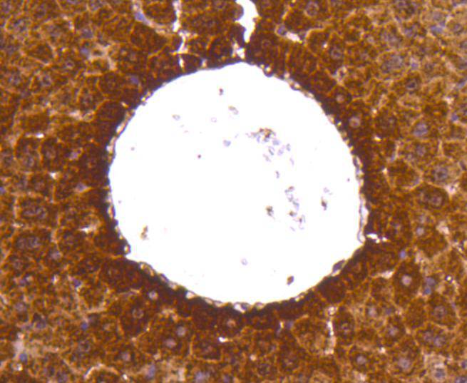
Immunohistochemical analysis of paraffin-embedded mouse liver tissue using anti-Lactate Dehydrogenase antibody. The section was pre-treated using heat mediated antigen retrieval with Tris-EDTA buffer (pH 9.0) for 20 minutes.The tissues were blocked in 5% BSA for 30 minutes at room temperature, washed with ddH2O and PBS, and then probed with the primary antibody (1/50) for 30 minutes at room temperature. The detection was performed using an HRP conjugated compact polymer system. DAB was used as the chromogen. Tissues were counterstained with hematoxylin and mounted with DPX.FC
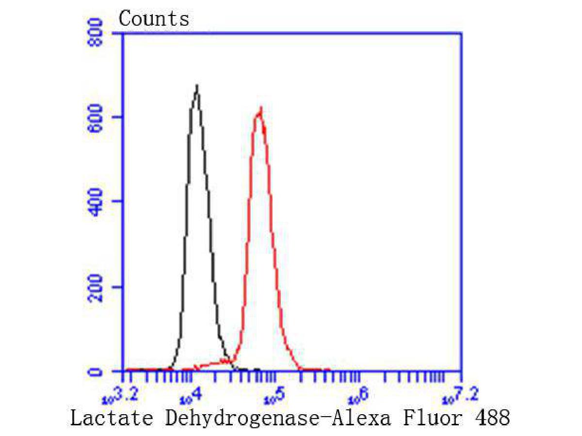
Flow cytometric analysis of Lactate Dehydrogenase was done on Hela cells. The cells were fixed, permeabilized and stained with the primary antibody (1/50) (red). After incubation of the primary antibody at room temperature for an hour, the cells were stained with a Alexa Fluor®488 conjugate-Goat anti-Rabbit IgG Secondary antibody at 1/1000 dilution for 30 minutes.Unlabelled sample was used as a control (cells without incubation with primary antibody; black).| Product Name | Lactate Dehydrogenase Recombinant Rabbit Monoclonal Antibody |
|---|---|
| Antibody Type | Primary Antibodies |
| Immunogen | Synthetic peptide within human LDHB aa 270-310. |
| Clonality | Monoclonal |
|---|---|
| Isotype | IgG |
| Host Species | Rabbit |
| Tested Applications | FCICC/IFIHCWB |
| WB:1:1000-1:5000 ICC/IF:1:50-1:200 IHC:1:200-1:2000 FC:1:50-1:100 |
|
| Species Reactivity | HumanMouse |
| Concentration | 1mg/ml |
| Purification | Affinity purified |
| Gene Symbol | LDHB |
|---|---|
| Gene Synonyms | LDH-B LDH-H LDHBD TRG-5 HEL-S-281 |
| Gene Full Name | lactate dehydrogenase B |
| Gene Summary | This gene encodes the B subunit of lactate dehydrogenase enzyme, which catalyzes the interconversion of pyruvate and lactate with concomitant interconversion of NADH and NAD+ in a post-glycolysis process. Alternatively spliced transcript variants have been found for this gene. Recent studies have shown that a C-terminally extended isoform is produced by use of an alternative in-frame translation termination codon via a stop codon readthrough mechanism, and that this isoform is localized in the peroxisomes. Mutations in this gene are associated with lactate dehydrogenase B deficiency. Pseudogenes have been identified on chromosomes X, 5 and 13. [provided by RefSeq, Feb 2016] |
| Alternative Names | Cell proliferation-inducing gene 19 protein antibody GSD11 antibody L lactate dehydrogenase B chain antibody L-lactate dehydrogenase A chain antibody Lactate dehydrogenase A antibody Lactate dehydrogenase B antibody Lactate dehydrogenase H chain antibody Lactate dehydrogenase M antibody LDH A antibody LDH B antibody Cell proliferation-inducing gene 19 protein antibody GSD11 antibody L lactate dehydrogenase B chain antibody L-lactate dehydrogenase A chain antibody Lactate dehydrogenase A antibody Lactate dehydrogenase B antibody Lactate dehydrogenase H chain antibody Lactate dehydrogenase M antibody LDH A antibody LDH B antibody LDH H antibody LDH heart subunit antibody LDH M antibody LDH muscle subunit antibody LDH-A antibody LDH-M antibody LDH1 antibody ldha antibody LDHA_HUMAN antibody LDHBD antibody LDHM antibody MS1111 antibody PIG19 antibody Proliferation inducing gene 19 antibody Renal carcinoma antigen NY REN 46 antibody Renal carcinoma antigen NY-REN-59 antibody TRG 5 antibody TRG5 antibody |
| Molecular Weight(MW) | 37kDa(Observed band size:37kDa) |
| Cellular Localization | Cytoplasm. |
WB
Western blot analysis of Lactate Dehydrogenase on different lysates with Rabbit anti-Lactate Dehydrogenase antibody at 1/5000 dilution. Lane 1: HeLa cell lysate (15 µg/Lane), Lane 2: HEK-293 cell lysate (15 µg/Lane), Lane 3: A549 cell lysate (15 µg/Lane), Exposure time: 1 minute 36 seconds; 4-20% SDS-PAGE gel. Proteins were transferred to a PVDF membrane and blocked with 5% NFDM/TBST for 1 hour at room temperature. The primary antibody at 1/5000 dilution was used in 5% NFDM/TBST at 4℃ overnight. Goat Anti-Rabbit IgG - HRP Secondary Antibody at 1:50000 dilution was used for 1 hour at room temperature.
WB
Western blot analysis of Lactate Dehydrogenase on different lysates with Rabbit anti-Lactate Dehydrogenase antibody at 1/1000 dilution. Lane 1: Hela-si NT cell lysate (10 µg/Lane), Lane 2: Hela-si Lactate Dehydrogenase cell lysate (10 µg/Lane), Exposure time: 25 seconds; 4-20% SDS-PAGE gel. Rabbit anti-Lactate Dehydrogenase antibody was shown to specifically react with Lactate Dehydrogenase in Hela-si NT cells. Weakened band was observed when Hela-si Lactate Dehydrogenase sample was tested. Hela-si NT and Hela-si Lactate Dehydrogenase samples were subjected to SDS-PAGE. Proteins were transferred to a PVDF membrane and blocked with 5% NFDM in TBST for 1 hour at room temperature. The primary antibody (1/1000) and Loading control antibody (Rabbit anti-GAPDH, 1/10000) were used in 5% BSA at room temperature for 2 hours. Goat Anti-rabbit IgG-HRP Secondary Antibody at 1:100000 dilution was used for 1 hour at room temperature.
WB
Western blot analysis of Lactate Dehydrogenase on hybrid fish (crucian-carp) brain tissue lysates. Proteins were transferred to a PVDF membrane and blocked with 5% BSA in PBS for 1 hour at room temperature. The primary antibody (1/500) was used in 5% BSA at room temperature for 2 hours. Goat Anti-Rabbit IgG - HRP Secondary Antibody at 1:200000 dilution was used for 1 hour at room temperature.
ICC/IF
ICC staining of Lactate Dehydrogenase in A549 cells (green). Formalin fixed cells were permeabilized with 0.1% Triton X-100 in TBS for 10 minutes at room temperature and blocked with 10% negative goat serum for 15 minutes at room temperature. Cells were probed with the primary antibody (1/50) for 1 hour at room temperature, washed with PBS. Alexa Fluor®488 conjugate-Goat anti-Rabbit IgG was used as the secondary antibody at 1/1000 dilution. The nuclear counter stain is DAPI (blue).
ICC/IF
ICC staining of Lactate Dehydrogenase in A431 cells (green). Formalin fixed cells were permeabilized with 0.1% Triton X-100 in TBS for 10 minutes at room temperature and blocked with 10% negative goat serum for 15 minutes at room temperature. Cells were probed with the primary antibody (1/50) for 1 hour at room temperature, washed with PBS. Alexa Fluor®488 conjugate-Goat anti-Rabbit IgG was used as the secondary antibody at 1/1000 dilution. The nuclear counter stain is DAPI (blue).
ICC/IF
Immunocytochemistry analysis of LOVO cells labeling Lactate Dehydrogenase with Rabbit anti-Lactate Dehydrogenase antibody at 1/50 dilution. Cells were fixed in 4% paraformaldehyde for 10 minutes at 37 ℃, permeabilized with 0.05% Triton X-100 in PBS for 20 minutes, and then blocked with 2% negative goat serum for 30 minutes at room temperature. Cells were then incubated with Rabbit anti-Lactate Dehydrogenase antibody at 1/50 dilution in 2% negative goat serum overnight at 4 ℃. Goat Anti-Rabbit IgG H&L (488) was used as the secondary antibody at 1/1,000 dilution. PBS instead of the primary antibody was used as the secondary antibody only control. Nuclear DNA was labelled in blue with DAPI.
IHC
Immunohistochemical analysis of paraffin-embedded human liver tissue using anti-Lactate Dehydrogenase antibody. The section was pre-treated using heat mediated antigen retrieval with Tris-EDTA buffer (pH 9.0) for 20 minutes.The tissues were blocked in 5% BSA for 30 minutes at room temperature, washed with ddH2O and PBS, and then probed with the primary antibody (1/50) for 30 minutes at room temperature. The detection was performed using an HRP conjugated compact polymer system. DAB was used as the chromogen. Tissues were counterstained with hematoxylin and mounted with DPX.
IHC
Immunohistochemical analysis of paraffin-embedded mouse liver tissue using anti-Lactate Dehydrogenase antibody. The section was pre-treated using heat mediated antigen retrieval with Tris-EDTA buffer (pH 9.0) for 20 minutes.The tissues were blocked in 5% BSA for 30 minutes at room temperature, washed with ddH2O and PBS, and then probed with the primary antibody (1/50) for 30 minutes at room temperature. The detection was performed using an HRP conjugated compact polymer system. DAB was used as the chromogen. Tissues were counterstained with hematoxylin and mounted with DPX.
FC
Flow cytometric analysis of Lactate Dehydrogenase was done on Hela cells. The cells were fixed, permeabilized and stained with the primary antibody (1/50) (red). After incubation of the primary antibody at room temperature for an hour, the cells were stained with a Alexa Fluor®488 conjugate-Goat anti-Rabbit IgG Secondary antibody at 1/1000 dilution for 30 minutes.Unlabelled sample was used as a control (cells without incubation with primary antibody; black).| Application Notes | WB:1:1000-1:5000 ICC/IF:1:50-1:200 IHC:1:200-1:2000 FC:1:50-1:100 |
|---|
| Form | Liquid |
|---|---|
| Storage Instructions | Shipped at 4℃. Store at +4℃ short term (1-2 weeks). It is recommended to aliquot into single-use upon delivery. Store at -20℃ long term. |
| Storage Buffer | 1*TBS (pH7.4), 0.05% BSA, 40% Glycerol. Preservative: 0.05% Sodium Azide. |


