WB
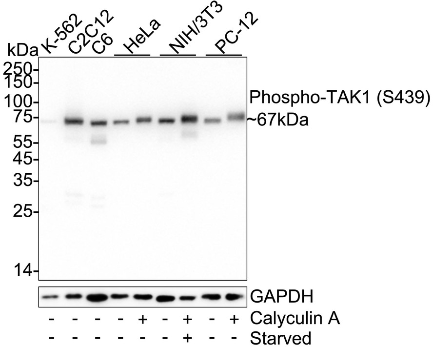
Western blot analysis of Phospho-TAK1 (S439) on different lysates with Rabbit anti-Phospho-TAK1 (S439) antibody at 1/25000 dilution. Lane 1: K-562 cell lysate, Lane 2: C2C12 cell lysate, Lane 3: C6 cell lysate, Lane 4: HeLa cell lysate, Lane 5: HeLa treated with 100nM Calyculin A for 30 minutes cell lysate, Lane 6: NIH/3T3 cell lysate, Lane 7: NIH/3T3 starved overnight then treated with 100nM Calyculin A for 30 minutes cell lysate, Lane 8: PC-12 cell lysate, Lane 9: PC-12 treated with 100nM Calyculin A for 30 minutes cell lysate, Lysates/proteins at 20 µg/Lane. Exposure time: 10 seconds; 4-20% SDS-PAGE gel. Proteins were transferred to a PVDF membrane and blocked with 5% NFDM/TBST for 1 hour at room temperature. The primary antibody at 1/25000 dilution was used in primary antibody dilution at 4℃ overnight. Goat Anti-Rabbit IgG - HRP Secondary Antibody at 1/50000 dilution was used for 1 hour at room temperature.WB
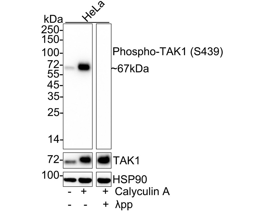
Western blot analysis of Phospho-TAK1 (S439) on different lysates with Rabbit anti-Phospho-TAK1 (S439) antibody at 1/5000 dilution and pan TAK1 antibody at 1/1000 dilution. Lane 1: HeLa cell lysate Lane 2: HeLa treated with 100ng/mL Calyculin A for 30 minutes cell lysate Lane 3: HeLa treated with 100ng/mL Calyculin A for 30 minutes cell lysate, then the membrane treated with λpp for 1 hour Lysates/proteins at 20 µg/Lane. Exposure time: 25 seconds; 4-20% SDS-PAGE gel. Proteins were transferred to a PVDF membrane and blocked with 5% NFDM/TBST for 1 hour at room temperature. The primary antibody at 1/5000 dilution and pan TAK1 antibody at 1/1000 dilution were used in 5% NFDM/TBST at 4℃ overnight. Goat Anti-Rabbit IgG - HRP Secondary Antibody at 1/50000 dilution was used for 1 hour at room temperature.| Product Name | Phospho-TAK1 (S439) Recombinant Rabbit Monoclonal Antibody |
|---|---|
| Antibody Type | Primary Antibodies |
| Immunogen | Synthetic phosphopeptide corresponding to residues surrounding Ser439 of mouse TAK1. |
| Modification | P-S439 |
| Clonality | Monoclonal |
|---|---|
| Isotype | IgG |
| Host Species | Rabbit |
| Tested Applications | WB |
| WB:1:5000-1:25000 |
|
| Species Reactivity | HumanMouseRat |
| Concentration | 1mg/ml |
| Purification | Affinity purified |
| Gene Symbol | MAP3K7 |
|---|---|
| Gene Synonyms | CSCF FMD2 TAK1 MEKK7 TGF1a |
| Gene Full Name | mitogen-activated protein kinase kinase kinase 7 |
| Gene Summary | The protein encoded by this gene is a member of the serine/threonine protein kinase family. This kinase mediates the signaling transduction induced by TGF beta and morphogenetic protein (BMP), and controls a variety of cell functions including transcription regulation and apoptosis. In response to IL-1, this protein forms a kinase complex including TRAF6, MAP3K7P1/TAB1 and MAP3K7P2/TAB2; this complex is required for the activation of nuclear factor kappa B. This kinase can also activate MAPK8/JNK, MAP2K4/MKK4, and thus plays a role in the cell response to environmental stresses. Four alternatively spliced transcript variants encoding distinct isoforms have been reported. [provided by RefSeq, Jul 2008] |
| Alternative Names | M3K7_HUMAN antibody MAP3K 7 antibody Map3k7 antibody MEKK7 antibody Mitogen activated protein kinase kinase kinase 7 antibody Mitogen-activated protein kinase kinase kinase 7 antibody TAK1 antibody TGF beta activated kinase 1 antibody TGF-beta-activated kinase 1 antibody TGF1a antibody M3K7_HUMAN antibody MAP3K 7 antibody Map3k7 antibody MEKK7 antibody Mitogen activated protein kinase kinase kinase 7 antibody Mitogen-activated protein kinase kinase kinase 7 antibody TAK1 antibody TGF beta activated kinase 1 antibody TGF-beta-activated kinase 1 antibody TGF1a antibody Transforming growth factor beta activated kinase 1 antibody Transforming growth factor-beta-activated kinase 1 antibody |
| Molecular Weight(MW) | 67kDa(Observed band size:67kDa) |
| Cellular Localization | Cytoplasm, Cell membrane. |
WB
Western blot analysis of Phospho-TAK1 (S439) on different lysates with Rabbit anti-Phospho-TAK1 (S439) antibody at 1/25000 dilution. Lane 1: K-562 cell lysate, Lane 2: C2C12 cell lysate, Lane 3: C6 cell lysate, Lane 4: HeLa cell lysate, Lane 5: HeLa treated with 100nM Calyculin A for 30 minutes cell lysate, Lane 6: NIH/3T3 cell lysate, Lane 7: NIH/3T3 starved overnight then treated with 100nM Calyculin A for 30 minutes cell lysate, Lane 8: PC-12 cell lysate, Lane 9: PC-12 treated with 100nM Calyculin A for 30 minutes cell lysate, Lysates/proteins at 20 µg/Lane. Exposure time: 10 seconds; 4-20% SDS-PAGE gel. Proteins were transferred to a PVDF membrane and blocked with 5% NFDM/TBST for 1 hour at room temperature. The primary antibody at 1/25000 dilution was used in primary antibody dilution at 4℃ overnight. Goat Anti-Rabbit IgG - HRP Secondary Antibody at 1/50000 dilution was used for 1 hour at room temperature.
WB
Western blot analysis of Phospho-TAK1 (S439) on different lysates with Rabbit anti-Phospho-TAK1 (S439) antibody at 1/5000 dilution and pan TAK1 antibody at 1/1000 dilution. Lane 1: HeLa cell lysate Lane 2: HeLa treated with 100ng/mL Calyculin A for 30 minutes cell lysate Lane 3: HeLa treated with 100ng/mL Calyculin A for 30 minutes cell lysate, then the membrane treated with λpp for 1 hour Lysates/proteins at 20 µg/Lane. Exposure time: 25 seconds; 4-20% SDS-PAGE gel. Proteins were transferred to a PVDF membrane and blocked with 5% NFDM/TBST for 1 hour at room temperature. The primary antibody at 1/5000 dilution and pan TAK1 antibody at 1/1000 dilution were used in 5% NFDM/TBST at 4℃ overnight. Goat Anti-Rabbit IgG - HRP Secondary Antibody at 1/50000 dilution was used for 1 hour at room temperature.| Application Notes | WB:1:5000-1:25000 |
|---|
| Form | Liquid |
|---|---|
| Storage Instructions | Shipped at 4℃. Store at +4℃ short term (1-2 weeks). It is recommended to aliquot into single-use upon delivery. Store at -20℃ long term. |
| Storage Buffer | 1*TBS (pH7.4), 0.05% BSA, 40% Glycerol. Preservative: 0.05% Sodium Azide. |


