WB
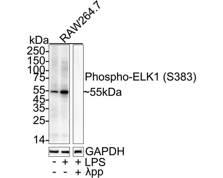
Western blot analysis of Phospho-ELK1 (S383) on different lysates with Rabbit anti-Phospho-ELK1 (S383) antibody at 1/5000 dilution. Lane 1: RAW264.7 cell lysate, Lane 2: RAW264.7 treated with 10μg/mL LPS for 8 hours cell lysate, Lane 3: RAW264.7 treated with 10μg/mL LPS for 8 hours cell lysate, then the membrane treated with λpp for 1 hour, Lysates/proteins at 20 µg/Lane. Exposure time: 59 seconds; 4-20% SDS-PAGE gel. Proteins were transferred to a PVDF membrane and blocked with 5% NFDM/TBST for 1 hour at room temperature. The primary antibody at 1/5000 dilution was used in primary antibody dilution at 4℃ overnight. Goat Anti-Rabbit IgG - HRP Secondary Antibody at 1/50000 dilution was used for 1 hour at room temperature.IHC
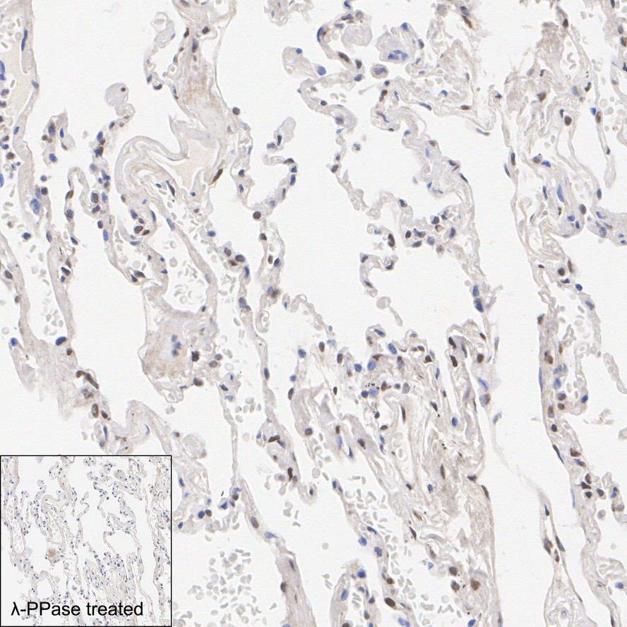
Immunohistochemical analysis of paraffin-embedded human lung tissue untreated / treated with λpp with Rabbit anti-Phospho-ELK1 (S383) antibody at 1/1000 dilution. The section was pre-treated using heat mediated antigen retrieval with Tris-EDTA buffer (pH 9.0) for 20 minutes. The tissues were blocked in 1% BSA for 20 minutes at room temperature, washed with ddH2O and PBS, and then probed with the primary antibody at 1/1000 dilution for 1 hour at room temperature. The detection was performed using an HRP conjugated compact polymer system. DAB was used as the chromogen. Tissues were counterstained with hematoxylin and mounted with DPX.IP
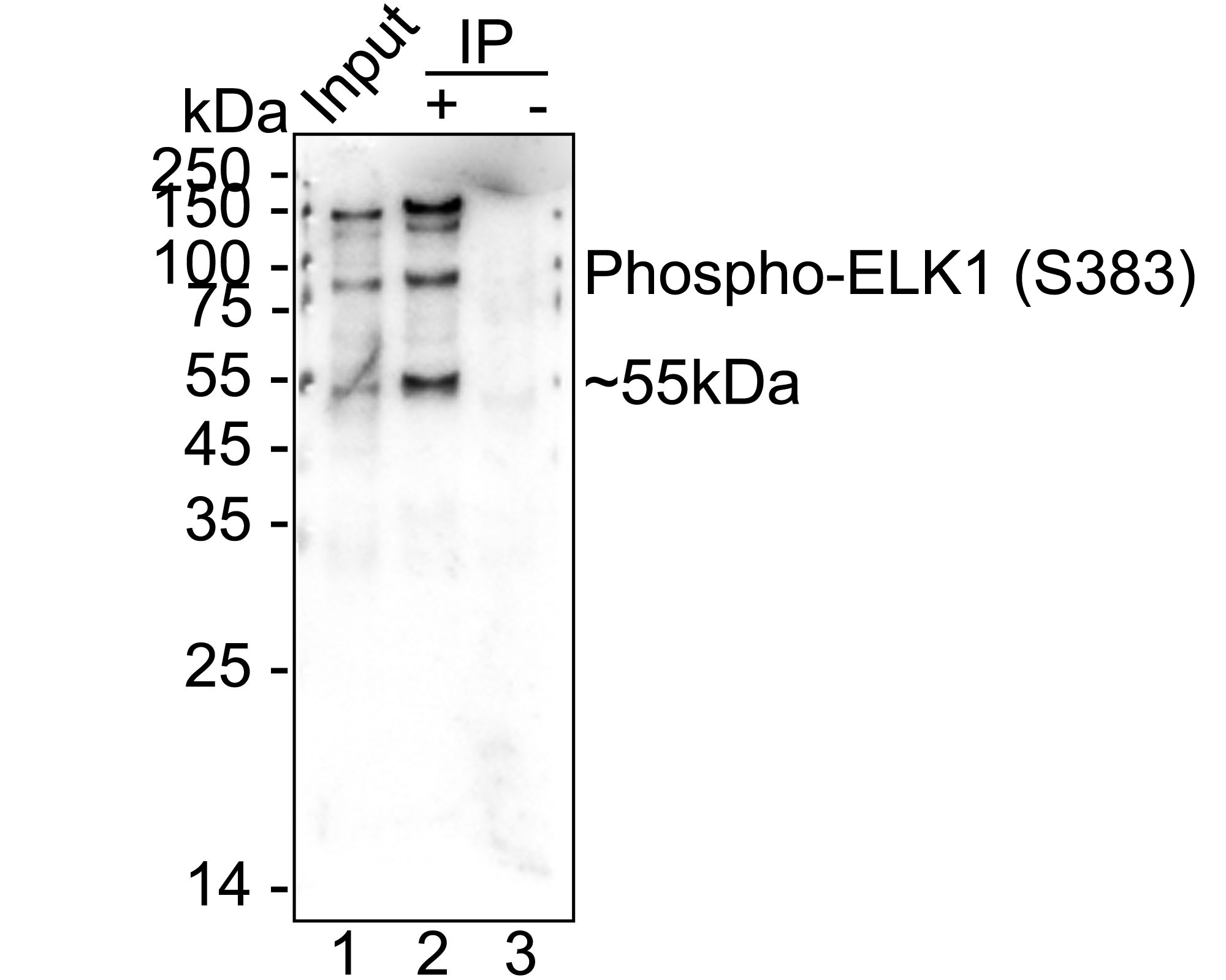
Phospho-ELK1 (S383) was immunoprecipitated from 0.2 mg RAW264.7 treated with 0.1μg/mL LPS for 6 hours cell lysate with Rabbit anti-Phospho-ELK1 (S383) antibody at 2 µg/10 µl beads. Western blot was performed from the immunoprecipitate using Rabbit anti-Phospho-ELK1 (S383) antibody at 1/25000 dilution. HRP Conjugated Anti-Rabbit IgG for IP Nano-secondary antibody at 1/5,000 dilution was used for 1 hour at room temperature. Lane 1: RAW264.7 treated with 0.1μg/mL LPS for 6 hours cell lysate (input), Lane 2: Rabbit anti-Phospho-ELK1 (S383) antibody IP in RAW264.7 treated with 0.1μg/mL LPS for 6 hours cell lysate, Lane 3: Rabbit IgG instead of Rabbit anti-Phospho-ELK1 (S383) antibody in RAW264.7 treated with 0.1μg/mL LPS for 6 hours cell lysate, Blocking/Dilution buffer: 5% NFDM/TBST, Exposure time: 1 minute 2 seconds.| Product Name | Phospho-ELK1 (S383) Recombinant Rabbit Monoclonal Antibody |
|---|---|
| Antibody Type | Primary Antibodies |
| Immunogen | Synthetic phosphopeptide corresponding to residues surrounding Ser383 of human ELK1. |
| Modification | P-S383 |
| Clonality | Monoclonal |
|---|---|
| Isotype | IgG |
| Host Species | Rabbit |
| Tested Applications | IHCIPWB |
| WB:1:5000 IP:1-2μg/sample IHC:1:1000 |
|
| Species Reactivity | HumanMouse |
| Concentration | 1mg/ml |
| Purification | Affinity purified |
| Gene Symbol | ELK1 |
|---|---|
| Gene Full Name | ETS transcription factor ELK1 |
| Gene Summary | This gene is a member of the Ets family of transcription factors and of the ternary complex factor (TCF) subfamily. Proteins of the TCF subfamily form a ternary complex by binding to the the serum response factor and the serum response element in the promoter of the c-fos proto-oncogene. The protein encoded by this gene is a nuclear target for the ras-raf-MAPK signaling cascade. This gene produces multiple isoforms by using alternative translational start codons and by alternative splicing. Related pseudogenes have been identified on chromosomes 7 and 14. [provided by RefSeq, Mar 2012] |
| Alternative Names | ELK 1 antibody Elk1 antibody ELK1 member of ETS oncogene family antibody ELK1 protein antibody ELK1 ETS transcription factor antibody ELK1_HUMAN antibody ELK2 member of ETS oncogene family antibody ETS domain containing protein Elk 1 antibody ETS domain containing protein Elk1 antibody ETS domain protein Elk1 antibody ELK 1 antibody Elk1 antibody ELK1 member of ETS oncogene family antibody ELK1 protein antibody ELK1 ETS transcription factor antibody ELK1_HUMAN antibody ELK2 member of ETS oncogene family antibody ETS domain containing protein Elk 1 antibody ETS domain containing protein Elk1 antibody ETS domain protein Elk1 antibody ETS domain-containing protein Elk-1 antibody ETS like gene 1 antibody Member of ETS oncogene family antibody Oncogene Elk1 antibody Tyrosine kinase (ELK1) oncogene antibody |
| Molecular Weight(MW) | 45kDa(Observed band size:55kDa) |
| Cellular Localization | Nucleus. |
WB
Western blot analysis of Phospho-ELK1 (S383) on different lysates with Rabbit anti-Phospho-ELK1 (S383) antibody at 1/5000 dilution. Lane 1: RAW264.7 cell lysate, Lane 2: RAW264.7 treated with 10μg/mL LPS for 8 hours cell lysate, Lane 3: RAW264.7 treated with 10μg/mL LPS for 8 hours cell lysate, then the membrane treated with λpp for 1 hour, Lysates/proteins at 20 µg/Lane. Exposure time: 59 seconds; 4-20% SDS-PAGE gel. Proteins were transferred to a PVDF membrane and blocked with 5% NFDM/TBST for 1 hour at room temperature. The primary antibody at 1/5000 dilution was used in primary antibody dilution at 4℃ overnight. Goat Anti-Rabbit IgG - HRP Secondary Antibody at 1/50000 dilution was used for 1 hour at room temperature.
IHC
Immunohistochemical analysis of paraffin-embedded human lung tissue untreated / treated with λpp with Rabbit anti-Phospho-ELK1 (S383) antibody at 1/1000 dilution. The section was pre-treated using heat mediated antigen retrieval with Tris-EDTA buffer (pH 9.0) for 20 minutes. The tissues were blocked in 1% BSA for 20 minutes at room temperature, washed with ddH2O and PBS, and then probed with the primary antibody at 1/1000 dilution for 1 hour at room temperature. The detection was performed using an HRP conjugated compact polymer system. DAB was used as the chromogen. Tissues were counterstained with hematoxylin and mounted with DPX.
IP
Phospho-ELK1 (S383) was immunoprecipitated from 0.2 mg RAW264.7 treated with 0.1μg/mL LPS for 6 hours cell lysate with Rabbit anti-Phospho-ELK1 (S383) antibody at 2 µg/10 µl beads. Western blot was performed from the immunoprecipitate using Rabbit anti-Phospho-ELK1 (S383) antibody at 1/25000 dilution. HRP Conjugated Anti-Rabbit IgG for IP Nano-secondary antibody at 1/5,000 dilution was used for 1 hour at room temperature. Lane 1: RAW264.7 treated with 0.1μg/mL LPS for 6 hours cell lysate (input), Lane 2: Rabbit anti-Phospho-ELK1 (S383) antibody IP in RAW264.7 treated with 0.1μg/mL LPS for 6 hours cell lysate, Lane 3: Rabbit IgG instead of Rabbit anti-Phospho-ELK1 (S383) antibody in RAW264.7 treated with 0.1μg/mL LPS for 6 hours cell lysate, Blocking/Dilution buffer: 5% NFDM/TBST, Exposure time: 1 minute 2 seconds.| Application Notes | WB:1:5000 IP:1-2μg/sample IHC:1:1000 |
|---|
| Form | Liquid |
|---|---|
| Storage Instructions | Shipped at 4℃. Store at +4℃ short term (1-2 weeks). It is recommended to aliquot into single-use upon delivery. Store at -20℃ long term. |
| Storage Buffer | PBS (pH7.4), 0.1% BSA, 40% Glycerol. Preservative: 0.05% Sodium Azide. |


