WB
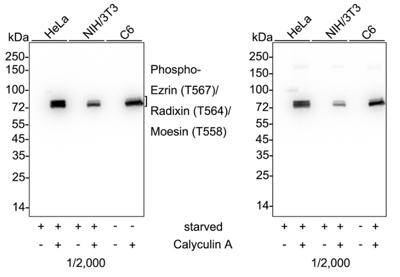
Western blot analysis of Phospho-Ezrin (T567)/Radixin (T564)/Moesin (T558) on different lysates with Rabbit anti-Phospho-Ezrin (T567)/Radixin (T564)/Moesin (T558) antibody at 1/2000 dilution and competitor's antibody at 1/2000 dilution. Lane 1: HeLa starved for 3 hours cell lysate, Lane 2: HeLa starved for 3 hours then add 100nM Calyculin A for 30 minutes cell lysate, Lane 3: NIH/3T3 starved for 24 hours cell lysate, Lane 4: NIH/3T3 starved for 24 hours then add 100nM Calyculin A for 30 minutes cell lysate, Lane 5: C6 cell lysate, Lane 6: C6 treated with 100ng/mL Calyculin A for 1 hours cell lysate, Lysates/proteins at 20 µg/Lane. Exposure time: 9 seconds; 4-20% SDS-PAGE gel. Proteins were transferred to a PVDF membrane and blocked with 5% NFDM/TBST for 1 hour at room temperature. The primary antibody at 1/2000 dilution and competitor's antibody at 1/2,000 dilution were used in 5% NFDM/TBST at 4℃ overnight. Goat Anti-Rabbit IgG - HRP Secondary Antibody at 1/50000 dilution was used for 1 hour at room temperature.ICC/IF
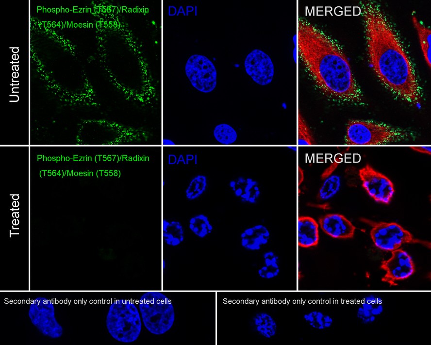
Immunocytochemistry analysis of HeLa cells untreated (upper, positive) and HeLa cells treated with 1μM staurosporine for 3 hours (lower, negative) labeling Phospho-Ezrin (T567)/Radixin (T564)/Moesin (T558) with Rabbit anti-Phospho-Ezrin (T567)/Radixin (T564)/Moesin (T558) antibody at 1/100 dilution. Cells were fixed in 4% paraformaldehyde for 20 minutes at room temperature, permeabilized with 0.1% Triton X-100 in PBS for 5 minutes at room temperature, then blocked with 1% BSA in 10% negative goat serum for 1 hour at room temperature. Cells were then incubated with Rabbit anti-Phospho-Ezrin (T567)/Radixin (T564)/Moesin (T558) antibody at 1/100 dilution in 1% BSA in PBST overnight at 4 ℃. Goat Anti-Rabbit IgG H&L (488) was used as the secondary antibody at 1/1,000 dilution. PBS instead of the primary antibody was used as the secondary antibody only control. Nuclear DNA was labelled in blue with DAPI. Beta tubulin (red) was stained at 1/100 dilution overnight at +4℃. Goat Anti-Mouse IgG H&L (594) was used as the secondary antibody at 1/1000 dilution.IHC
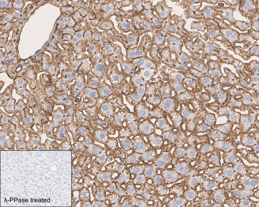
Immunohistochemical analysis of paraffin-embedded mouse liver tissue untreated / treated with λpp with Rabbit anti-Phospho-Ezrin (T567)/Radixin (T564)/Moesin (T558) antibody at 1/1000 dilution. The section was pre-treated using heat mediated antigen retrieval with Tris-EDTA buffer (pH 9.0) for 20 minutes. The tissues were blocked in 1% BSA for 20 minutes at room temperature, washed with ddH2O and PBS, and then probed with the primary antibody at 1/1000 dilution for 1 hour at room temperature. The detection was performed using an HRP conjugated compact polymer system. DAB was used as the chromogen. Tissues were counterstained with hematoxylin and mounted with DPX.IHC
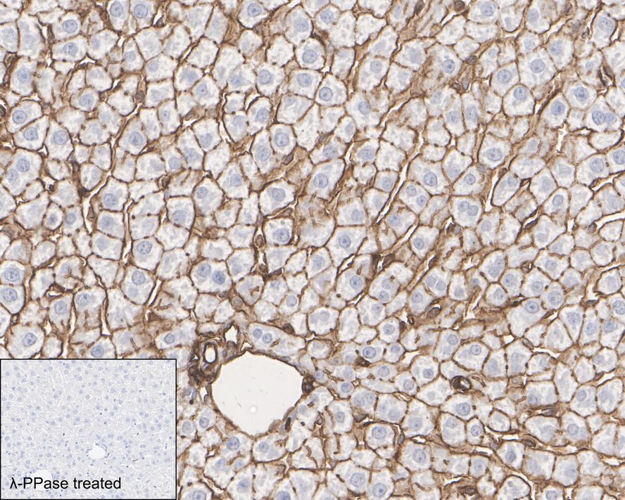
Immunohistochemical analysis of paraffin-embedded rat liver tissue untreated / treated with λpp with Rabbit anti-Phospho-Ezrin (T567)/Radixin (T564)/Moesin (T558) antibody at 1/1000 dilution. The section was pre-treated using heat mediated antigen retrieval with Tris-EDTA buffer (pH 9.0) for 20 minutes. The tissues were blocked in 1% BSA for 20 minutes at room temperature, washed with ddH2O and PBS, and then probed with the primary antibody at 1/1000 dilution for 1 hour at room temperature. The detection was performed using an HRP conjugated compact polymer system. DAB was used as the chromogen. Tissues were counterstained with hematoxylin and mounted with DPX.| Product Name | Phospho-Ezrin (T567)/Radixin (T564)/Moesin (T558) Recombinant Rabbit Monoclonal Antibody |
|---|---|
| Antibody Type | Primary Antibodies |
| Immunogen | Synthetic phosphopeptide corresponding to residues surrounding Thr567 of human ezrin protein. |
| Modification | P-T567 |
| Clonality | Monoclonal |
|---|---|
| Isotype | IgG |
| Host Species | Rabbit |
| Tested Applications | ICC/IFIHCWB |
| WB:1:2000 ICC/IF:1:100 IHC:1:1000 |
|
| Species Reactivity | HumanMouseRat |
| Concentration | 1mg/ml |
| Purification | Affinity purified |
| Gene Symbol | EZR |
|---|---|
| Gene Synonyms | CVL CVIL VIL2 HEL-S-105 |
| Gene Full Name | ezrin |
| Gene Summary | The cytoplasmic peripheral membrane protein encoded by this gene functions as a protein-tyrosine kinase substrate in microvilli. As a member of the ERM protein family, this protein serves as an intermediate between the plasma membrane and the actin cytoskeleton. This protein plays a key role in cell surface structure adhesion, migration and organization, and it has been implicated in various human cancers. A pseudogene located on chromosome 3 has been identified for this gene. Alternatively spliced variants have also been described for this gene. [provided by RefSeq, Jul 2008] |
| Alternative Names | Villin 2 ezrin antibody CVIL antibody CVL antibody Cytovillin 2 antibody Cytovillin antibody DKFZp762H157 antibody Epididymis secretory protein Li 105 antibody EZR antibody EZRI_HUMAN antibody Ezrin antibody Villin 2 ezrin antibody CVIL antibody CVL antibody Cytovillin 2 antibody Cytovillin antibody DKFZp762H157 antibody Epididymis secretory protein Li 105 antibody EZR antibody EZRI_HUMAN antibody Ezrin antibody FLJ26216 antibody HEL S 105 antibody MGC1584 antibody p81 antibody VIL 2 antibody VIL2 antibody Villin 2 (ezrin) antibody Villin 2 antibody Villin-2 antibody Villin2 antibody CB567 antibody CG12537 antibody DFNB24 antibody ESP10 antibody Hh-induced MATH and BTB domain-containing protein antibody HIB antibody Moesin-B antibody Protein roadkill antibody RADI_HUMAN antibody Radixin antibody RDX antibody Epididymis luminal protein 70 antibody HEL70 antibody Membrane organizing extension spike protein antibody Membrane-organizing extension spike protein antibody MOES_HUMAN antibody Moesin antibody Moesin/anaplastic lymphoma kinase fusion protein antibody Msn antibody MSN/ALK fusion antibody |
| Molecular Weight(MW) | 69kDa(Observed band size:75/80kDa) |
| Cellular Localization | Cell membrane. Cell projection. Cytoplasm. Cytoskeleton. Membrane |
WB
Western blot analysis of Phospho-Ezrin (T567)/Radixin (T564)/Moesin (T558) on different lysates with Rabbit anti-Phospho-Ezrin (T567)/Radixin (T564)/Moesin (T558) antibody at 1/2000 dilution and competitor's antibody at 1/2000 dilution. Lane 1: HeLa starved for 3 hours cell lysate, Lane 2: HeLa starved for 3 hours then add 100nM Calyculin A for 30 minutes cell lysate, Lane 3: NIH/3T3 starved for 24 hours cell lysate, Lane 4: NIH/3T3 starved for 24 hours then add 100nM Calyculin A for 30 minutes cell lysate, Lane 5: C6 cell lysate, Lane 6: C6 treated with 100ng/mL Calyculin A for 1 hours cell lysate, Lysates/proteins at 20 µg/Lane. Exposure time: 9 seconds; 4-20% SDS-PAGE gel. Proteins were transferred to a PVDF membrane and blocked with 5% NFDM/TBST for 1 hour at room temperature. The primary antibody at 1/2000 dilution and competitor's antibody at 1/2,000 dilution were used in 5% NFDM/TBST at 4℃ overnight. Goat Anti-Rabbit IgG - HRP Secondary Antibody at 1/50000 dilution was used for 1 hour at room temperature.
ICC/IF
Immunocytochemistry analysis of HeLa cells untreated (upper, positive) and HeLa cells treated with 1μM staurosporine for 3 hours (lower, negative) labeling Phospho-Ezrin (T567)/Radixin (T564)/Moesin (T558) with Rabbit anti-Phospho-Ezrin (T567)/Radixin (T564)/Moesin (T558) antibody at 1/100 dilution. Cells were fixed in 4% paraformaldehyde for 20 minutes at room temperature, permeabilized with 0.1% Triton X-100 in PBS for 5 minutes at room temperature, then blocked with 1% BSA in 10% negative goat serum for 1 hour at room temperature. Cells were then incubated with Rabbit anti-Phospho-Ezrin (T567)/Radixin (T564)/Moesin (T558) antibody at 1/100 dilution in 1% BSA in PBST overnight at 4 ℃. Goat Anti-Rabbit IgG H&L (488) was used as the secondary antibody at 1/1,000 dilution. PBS instead of the primary antibody was used as the secondary antibody only control. Nuclear DNA was labelled in blue with DAPI. Beta tubulin (red) was stained at 1/100 dilution overnight at +4℃. Goat Anti-Mouse IgG H&L (594) was used as the secondary antibody at 1/1000 dilution.
IHC
Immunohistochemical analysis of paraffin-embedded mouse liver tissue untreated / treated with λpp with Rabbit anti-Phospho-Ezrin (T567)/Radixin (T564)/Moesin (T558) antibody at 1/1000 dilution. The section was pre-treated using heat mediated antigen retrieval with Tris-EDTA buffer (pH 9.0) for 20 minutes. The tissues were blocked in 1% BSA for 20 minutes at room temperature, washed with ddH2O and PBS, and then probed with the primary antibody at 1/1000 dilution for 1 hour at room temperature. The detection was performed using an HRP conjugated compact polymer system. DAB was used as the chromogen. Tissues were counterstained with hematoxylin and mounted with DPX.
IHC
Immunohistochemical analysis of paraffin-embedded rat liver tissue untreated / treated with λpp with Rabbit anti-Phospho-Ezrin (T567)/Radixin (T564)/Moesin (T558) antibody at 1/1000 dilution. The section was pre-treated using heat mediated antigen retrieval with Tris-EDTA buffer (pH 9.0) for 20 minutes. The tissues were blocked in 1% BSA for 20 minutes at room temperature, washed with ddH2O and PBS, and then probed with the primary antibody at 1/1000 dilution for 1 hour at room temperature. The detection was performed using an HRP conjugated compact polymer system. DAB was used as the chromogen. Tissues were counterstained with hematoxylin and mounted with DPX.| Application Notes | WB:1:2000 ICC/IF:1:100 IHC:1:1000 |
|---|
| Form | Liquid |
|---|---|
| Storage Instructions | Shipped at 4℃. Store at +4℃ short term (1-2 weeks). It is recommended to aliquot into single-use upon delivery. Store at -20℃ long term. |
| Storage Buffer | PBS (pH7.4), 0.1% BSA, 40% Glycerol. Preservative: 0.05% Sodium Azide. |


