WB
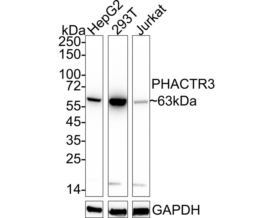
Western blot analysis of PHACTR3 on different lysates with Rabbit anti-PHACTR3 antibody at 1/1000 dilution. Lane 1: HepG2 cell lysate, Lane 2: 293T cell lysate, Lane 3: Jurkat cell lysate, Lysates/proteins at 20 µg/Lane. Exposure time: 25 seconds; 4-20% SDS-PAGE gel. Proteins were transferred to a PVDF membrane and blocked with 5% NFDM/TBST for 1 hour at room temperature. The primary antibody at 1/1000 dilution was used in 5% NFDM/TBST at 4℃ overnight. Goat Anti-Rabbit IgG - HRP Secondary Antibody at 1/50000 dilution was used for 1 hour at room temperature.ICC/IF
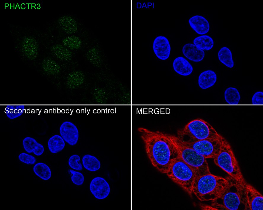
Immunocytochemistry analysis of HepG2 cells labeling PHACTR3 with Rabbit anti-PHACTR3 antibody at 1/50 dilution. Cells were fixed in 4% paraformaldehyde for 20 minutes at room temperature, permeabilized with 0.1% Triton X-100 in PBS for 5 minutes at room temperature, then blocked with 1% BSA in 10% negative goat serum for 1 hour at room temperature. Cells were then incubated with Rabbit anti-PHACTR3 antibody at 1/50 dilution in 1% BSA in PBST overnight at 4 ℃. Goat Anti-Rabbit IgG H&L (488) was used as the secondary antibody at 1/1,000 dilution. PBS instead of the primary antibody was used as the secondary antibody only control. Nuclear DNA was labelled in blue with DAPI. Beta tubulin (red) was stained at 1/100 dilution overnight at +4℃. Goat Anti-Mouse IgG H&L (594) was used as the secondary antibody at 1/1000 dilution.| Product Name | PHACTR3 Recombinant Rabbit Monoclonal Antibody |
|---|---|
| Antibody Type | Primary Antibodies |
| Immunogen | Synthetic peptide within human PHACTR3 aa 401-450. |
| Clonality | Monoclonal |
|---|---|
| Isotype | IgG |
| Host Species | Rabbit |
| Tested Applications | ICC/IFWB |
| WB:1:1000 ICC/IF:1:50 |
|
| Species Reactivity | Human |
| Concentration | 1mg/ml |
| Purification | Affinity purified |
| Gene Symbol | PHACTR3 |
|---|---|
| Gene Synonyms | H17739 SCAPIN1 PPP1R123 SCAPININ C20orf101 |
| Gene Full Name | phosphatase and actin regulator 3 |
| Gene Summary | This gene encodes a member of the phosphatase and actin regulator protein family. The encoded protein is associated with the nuclear scaffold in proliferating cells, and binds to actin and the catalytic subunit of protein phosphatase-1, suggesting that it functions as a regulatory subunit of protein phosphatase-1. Alternative splicing results in multiple transcript variants. [provided by RefSeq, Jul 2013] |
| Alternative Names | Phosphatase and actin regulator 3 Scaffold-associated PP1-inhibiting protein (Scapinin) PHACTR3 C20orf101 SCAPIN1 |
| Molecular Weight(MW) | 63kDa(Observed band size:63kDa) |
| Cellular Localization | Nucleus matrix. |
WB
Western blot analysis of PHACTR3 on different lysates with Rabbit anti-PHACTR3 antibody at 1/1000 dilution. Lane 1: HepG2 cell lysate, Lane 2: 293T cell lysate, Lane 3: Jurkat cell lysate, Lysates/proteins at 20 µg/Lane. Exposure time: 25 seconds; 4-20% SDS-PAGE gel. Proteins were transferred to a PVDF membrane and blocked with 5% NFDM/TBST for 1 hour at room temperature. The primary antibody at 1/1000 dilution was used in 5% NFDM/TBST at 4℃ overnight. Goat Anti-Rabbit IgG - HRP Secondary Antibody at 1/50000 dilution was used for 1 hour at room temperature.
ICC/IF
Immunocytochemistry analysis of HepG2 cells labeling PHACTR3 with Rabbit anti-PHACTR3 antibody at 1/50 dilution. Cells were fixed in 4% paraformaldehyde for 20 minutes at room temperature, permeabilized with 0.1% Triton X-100 in PBS for 5 minutes at room temperature, then blocked with 1% BSA in 10% negative goat serum for 1 hour at room temperature. Cells were then incubated with Rabbit anti-PHACTR3 antibody at 1/50 dilution in 1% BSA in PBST overnight at 4 ℃. Goat Anti-Rabbit IgG H&L (488) was used as the secondary antibody at 1/1,000 dilution. PBS instead of the primary antibody was used as the secondary antibody only control. Nuclear DNA was labelled in blue with DAPI. Beta tubulin (red) was stained at 1/100 dilution overnight at +4℃. Goat Anti-Mouse IgG H&L (594) was used as the secondary antibody at 1/1000 dilution.| Application Notes | WB:1:1000 ICC/IF:1:50 |
|---|
| Form | Liquid |
|---|---|
| Storage Instructions | Shipped at 4℃. Store at +4℃ short term (1-2 weeks). It is recommended to aliquot into single-use upon delivery. Store at -20℃ long term. |
| Storage Buffer | PBS (pH7.4), 0.1% BSA, 40% Glycerol. Preservative: 0.05% Sodium Azide. |


