WB
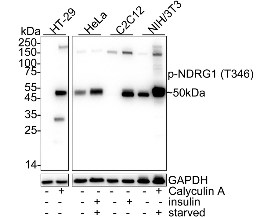
Western blot analysis of Phospho-NDRG1 (T346) on different lysates with Rabbit anti-Phospho-NDRG1 (T346) antibody at 1/2000 dilution. Lane 1: HT-29 cell lysate (25 µg/Lane), Lane 2: HT-29 treated with 100nM Calyculin A for 30 minutes cell lysate (25 µg/Lane), Lane 3: HeLa cell lysate (25 µg/Lane), Lane 4: HeLa serum starved for 4 hours then add 100nM insulin for 5 minutes cell lysate (25 µg/Lane), Lane 5: C2C12 cell lysate (25 µg/Lane), Lane 6: C2C12 treated with 100nM insulin for 15 minutes cell lysate (25 µg/Lane), Lane 7: HeLa cell lysate (25 µg/Lane), Lane 8: HeLa serum starved for 24 hours then add 100nM Calyculin A for 30 minutes cell lysate (25 µg/Lane), Exposure time: 20 seconds; 4-20% SDS-PAGE gel. Proteins were transferred to a PVDF membrane and blocked with 5% NFDM/TBST for 1 hour at room temperature. The primary antibody at 1/2000 dilution was used in 5% NFDM/TBST at 4℃ overnight. Goat Anti-Rabbit IgG - HRP Secondary Antibody at 1/50000 dilution was used for 1 hour at room temperature.WB
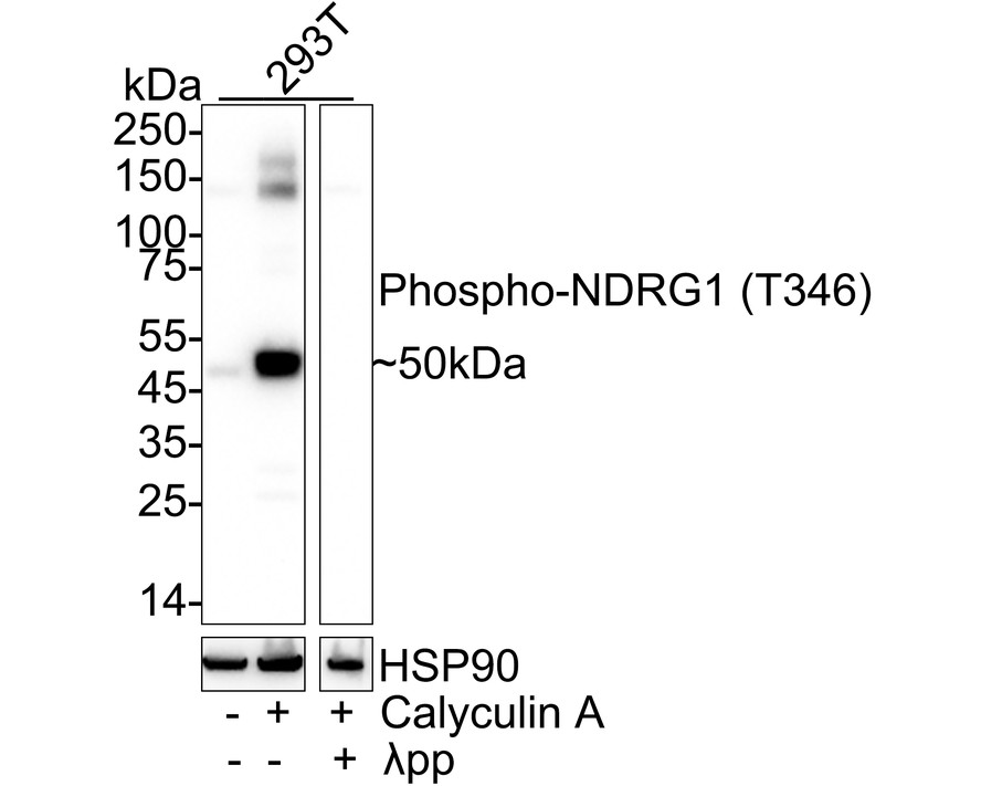
Western blot analysis of Phospho-NDRG1 (T346) on different lysates with Rabbit anti-Phospho-NDRG1 (T346) antibody at 1/2000 dilution. Lane 1: 293T cell lysate, Lane 2: 293T treated with 100nM Calyculin A for 15 minutes cell lysate, Lane 3: 293T treated with 100nM Calyculin A for 30 minutes cell lysate, then the membrane treated with λpp for 1 hour, Lysates/proteins at 20 µg/Lane. Exposure time: 10 seconds; 4-20% SDS-PAGE gel. Proteins were transferred to a PVDF membrane and blocked with 5% NFDM/TBST for 1 hour at room temperature. The primary antibody at 1/2000 dilution was used in 5% NFDM/TBST at 4℃ overnight. Goat Anti-Rabbit IgG - HRP Secondary Antibody at 1/50000 dilution was used for 1 hour at room temperature.WB
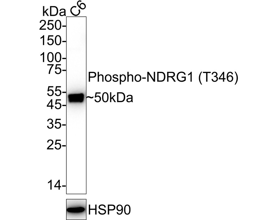
Western blot analysis of Phospho-NDRG1 (T346) on C6 cell lysates with Rabbit anti-Phospho-NDRG1 (T346) antibody at 1/2000 dilution. Lysates/proteins at 20 µg/Lane. Exposure time: 30 seconds; 4-20% SDS-PAGE gel. Proteins were transferred to a PVDF membrane and blocked with 5% NFDM/TBST for 1 hour at room temperature. The primary antibody at 1/2000 dilution was used in 5% NFDM/TBST at 4℃ overnight. Goat Anti-Rabbit IgG - HRP Secondary Antibody at 1/50000 dilution was used for 1 hour at room temperature.ICC/IF
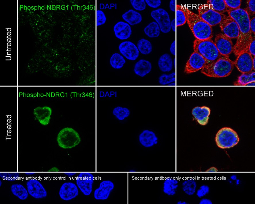
Immunocytochemistry analysis of 293T cells treated with 100nM Calyculin A for 15 minutes labeling Phospho-NDRG1 (T346) with Rabbit anti-Phospho-NDRG1 (T346) antibody at 1/100 dilution. Cells were fixed in 4% paraformaldehyde for 20 minutes at room temperature, permeabilized with 0.1% Triton X-100 in PBS for 5 minutes at room temperature, then blocked with 1% BSA in 10% negative goat serum for 1 hour at room temperature. Cells were then incubated with Rabbit anti-Phospho-NDRG1 (T346) antibody at 1/100 dilution in 1% BSA in PBST overnight at 4 ℃. Goat Anti-Rabbit IgG H&L (488) was used as the secondary antibody at 1/1000 dilution. PBS instead of the primary antibody was used as the secondary antibody only control. Nuclear DNA was labelled in blue with DAPI. Beta tubulin (red) was stained at 1/100 dilution overnight at +4℃. Goat Anti-Mouse IgG H&L (594) was used as the secondary antibody at 1/1000 dilution.IHC
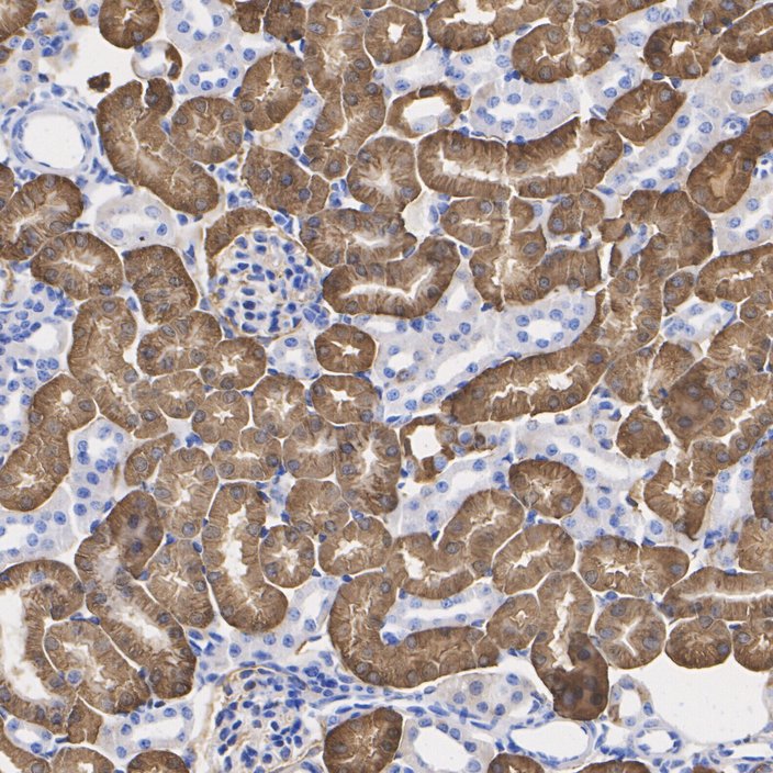
Immunohistochemical analysis of paraffin-embedded mouse kidney tissue with Rabbit anti-Phospho-NDRG1 (T346) antibody at 1/2000 dilution. The section was pre-treated using heat mediated antigen retrieval with Tris-EDTA buffer (pH 9.0) for 20 minutes. The tissues were blocked in 1% BSA for 20 minutes at room temperature, washed with ddH2O and PBS, and then probed with the primary antibody at 1/2000 dilution for 1 hour at room temperature. The detection was performed using an HRP conjugated compact polymer system. DAB was used as the chromogen. Tissues were counterstained with hematoxylin and mounted with DPX.IHC
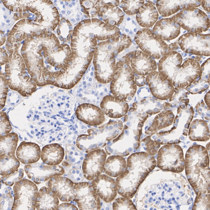
Immunohistochemical analysis of paraffin-embedded rat kidney tissue with Rabbit anti-Phospho-NDRG1 (T346) antibody at 1/2000 dilution. The section was pre-treated using heat mediated antigen retrieval with Tris-EDTA buffer (pH 9.0) for 20 minutes. The tissues were blocked in 1% BSA for 20 minutes at room temperature, washed with ddH2O and PBS, and then probed with the primary antibody at 1/2000 dilution for 1 hour at room temperature. The detection was performed using an HRP conjugated compact polymer system. DAB was used as the chromogen. Tissues were counterstained with hematoxylin and mounted with DPX.FC
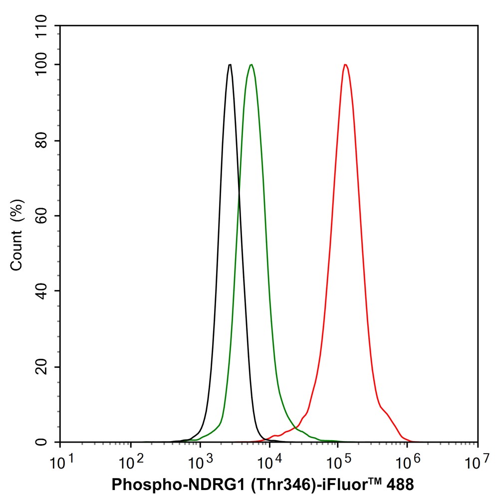
Flow cytometric analysis of 293T cells labeling Phospho-NDRG1 (T346). Cells were fixed and permeabilized. Then stained with the primary antibody (1/1000) (red) compared with Rabbit IgG Isotype Control (green). After incubation of the primary antibody at +4℃ for an hour, the cells were stained with a iFluor™ 488 conjugate-Goat anti-Rabbit IgG Secondary antibody at 1/1000 dilution for 30 minutes at +4℃. Unlabelled sample was used as a control (cells without incubation with primary antibody; black).| Product Name | Phospho-NDRG1 (T346) Recombinant Rabbit Monoclonal Antibody |
|---|---|
| Antibody Type | Primary Antibodies |
| Immunogen | Recombinant Rabbit monoclonal Antibody |
| Modification | P-T346 |
| Clonality | Monoclonal |
|---|---|
| Isotype | IgG |
| Host Species | Rabbit |
| Tested Applications | FCICC/IFIHCWB |
| WB:1:2000 IHC:1:2000 FC:1:1000 ICC/IF:1:100 |
|
| Species Reactivity | HumanMouseRat |
| Concentration | 1mg/ml |
| Purification | Affinity purified |
| Gene Symbol | NDRG1 |
|---|---|
| Gene Synonyms | GC4 RTP DRG1 NDR1 NMSL TDD5 CAP43 CMT4D DRG-1 HMSNL RIT42 TARG1 PROXY1 |
| Gene Full Name | N-myc downstream regulated 1 |
| Gene Summary | This gene is a member of the N-myc downregulated gene family which belongs to the alpha/beta hydrolase superfamily. The protein encoded by this gene is a cytoplasmic protein involved in stress responses, hormone responses, cell growth, and differentiation. The encoded protein is necessary for p53-mediated caspase activation and apoptosis. Mutations in this gene are a cause of Charcot-Marie-Tooth disease type 4D, and expression of this gene may be a prognostic indicator for several types of cancer. Alternatively spliced transcript variants encoding multiple isoforms have been observed for this gene. [provided by RefSeq, May 2012] |
| Alternative Names | 42 kDa antibody Anti GC4 antibody cap43 antibody cmt4d antibody Differentiation related gene1 protein antibody Differentiation-related gene 1 protein antibody Drg 1 antibody DRG-1 antibody drg1 antibody gc4 antibody 42 kDa antibody Anti GC4 antibody cap43 antibody cmt4d antibody Differentiation related gene1 protein antibody Differentiation-related gene 1 protein antibody Drg 1 antibody DRG-1 antibody drg1 antibody gc4 antibody GC4 antibody antibody hmsnl antibody Human mRNA for RTP complete cds antibody N myc downstream regulated gene 1 antibody N myc downstream regulated gene 1 protein antibody N-myc downstream-regulated gene 1 protein antibody Ndr 1 antibody ndr1 antibody NDRG 1 antibody Ndrg1 antibody NDRG1 protein antibody NDRG1_HUMAN antibody Nickel specific induction protein antibody Nickel specific induction protein Cap43 antibody Nickel-specific induction protein Cap43 antibody nmsl antibody Nmyc downstream regulated antibody Nmyc downstream regulated gene1 antibody Nmyc downstream regulated gene1 protein antibody Protein NDRG1 antibody Protein regulated by oxygen 1 antibody Protein regulated by oxygen1 antibody Proxy1 antibody Reduced in tumor antibody Reducin antibody Reducing agents and tunicamycin responsive protein antibody Reducing agents and tunicamycin-responsive protein antibody Rit42 antibody RTP antibody targ1 antibody TDD5 antibody tdds antibody Tunicamycin responsive protein antibody |
| Molecular Weight(MW) | 43kDa(Observed band size:50kDa) |
| Cellular Localization | Cytoplasm, cytosol, cytoskeleton, microtubule organizing center, centrosome, Nucleus, Cell membrane. |
WB
Western blot analysis of Phospho-NDRG1 (T346) on different lysates with Rabbit anti-Phospho-NDRG1 (T346) antibody at 1/2000 dilution. Lane 1: HT-29 cell lysate (25 µg/Lane), Lane 2: HT-29 treated with 100nM Calyculin A for 30 minutes cell lysate (25 µg/Lane), Lane 3: HeLa cell lysate (25 µg/Lane), Lane 4: HeLa serum starved for 4 hours then add 100nM insulin for 5 minutes cell lysate (25 µg/Lane), Lane 5: C2C12 cell lysate (25 µg/Lane), Lane 6: C2C12 treated with 100nM insulin for 15 minutes cell lysate (25 µg/Lane), Lane 7: HeLa cell lysate (25 µg/Lane), Lane 8: HeLa serum starved for 24 hours then add 100nM Calyculin A for 30 minutes cell lysate (25 µg/Lane), Exposure time: 20 seconds; 4-20% SDS-PAGE gel. Proteins were transferred to a PVDF membrane and blocked with 5% NFDM/TBST for 1 hour at room temperature. The primary antibody at 1/2000 dilution was used in 5% NFDM/TBST at 4℃ overnight. Goat Anti-Rabbit IgG - HRP Secondary Antibody at 1/50000 dilution was used for 1 hour at room temperature.
WB
Western blot analysis of Phospho-NDRG1 (T346) on different lysates with Rabbit anti-Phospho-NDRG1 (T346) antibody at 1/2000 dilution. Lane 1: 293T cell lysate, Lane 2: 293T treated with 100nM Calyculin A for 15 minutes cell lysate, Lane 3: 293T treated with 100nM Calyculin A for 30 minutes cell lysate, then the membrane treated with λpp for 1 hour, Lysates/proteins at 20 µg/Lane. Exposure time: 10 seconds; 4-20% SDS-PAGE gel. Proteins were transferred to a PVDF membrane and blocked with 5% NFDM/TBST for 1 hour at room temperature. The primary antibody at 1/2000 dilution was used in 5% NFDM/TBST at 4℃ overnight. Goat Anti-Rabbit IgG - HRP Secondary Antibody at 1/50000 dilution was used for 1 hour at room temperature.
WB
Western blot analysis of Phospho-NDRG1 (T346) on C6 cell lysates with Rabbit anti-Phospho-NDRG1 (T346) antibody at 1/2000 dilution. Lysates/proteins at 20 µg/Lane. Exposure time: 30 seconds; 4-20% SDS-PAGE gel. Proteins were transferred to a PVDF membrane and blocked with 5% NFDM/TBST for 1 hour at room temperature. The primary antibody at 1/2000 dilution was used in 5% NFDM/TBST at 4℃ overnight. Goat Anti-Rabbit IgG - HRP Secondary Antibody at 1/50000 dilution was used for 1 hour at room temperature.
ICC/IF
Immunocytochemistry analysis of 293T cells treated with 100nM Calyculin A for 15 minutes labeling Phospho-NDRG1 (T346) with Rabbit anti-Phospho-NDRG1 (T346) antibody at 1/100 dilution. Cells were fixed in 4% paraformaldehyde for 20 minutes at room temperature, permeabilized with 0.1% Triton X-100 in PBS for 5 minutes at room temperature, then blocked with 1% BSA in 10% negative goat serum for 1 hour at room temperature. Cells were then incubated with Rabbit anti-Phospho-NDRG1 (T346) antibody at 1/100 dilution in 1% BSA in PBST overnight at 4 ℃. Goat Anti-Rabbit IgG H&L (488) was used as the secondary antibody at 1/1000 dilution. PBS instead of the primary antibody was used as the secondary antibody only control. Nuclear DNA was labelled in blue with DAPI. Beta tubulin (red) was stained at 1/100 dilution overnight at +4℃. Goat Anti-Mouse IgG H&L (594) was used as the secondary antibody at 1/1000 dilution.
IHC
Immunohistochemical analysis of paraffin-embedded mouse kidney tissue with Rabbit anti-Phospho-NDRG1 (T346) antibody at 1/2000 dilution. The section was pre-treated using heat mediated antigen retrieval with Tris-EDTA buffer (pH 9.0) for 20 minutes. The tissues were blocked in 1% BSA for 20 minutes at room temperature, washed with ddH2O and PBS, and then probed with the primary antibody at 1/2000 dilution for 1 hour at room temperature. The detection was performed using an HRP conjugated compact polymer system. DAB was used as the chromogen. Tissues were counterstained with hematoxylin and mounted with DPX.
IHC
Immunohistochemical analysis of paraffin-embedded rat kidney tissue with Rabbit anti-Phospho-NDRG1 (T346) antibody at 1/2000 dilution. The section was pre-treated using heat mediated antigen retrieval with Tris-EDTA buffer (pH 9.0) for 20 minutes. The tissues were blocked in 1% BSA for 20 minutes at room temperature, washed with ddH2O and PBS, and then probed with the primary antibody at 1/2000 dilution for 1 hour at room temperature. The detection was performed using an HRP conjugated compact polymer system. DAB was used as the chromogen. Tissues were counterstained with hematoxylin and mounted with DPX.
FC
Flow cytometric analysis of 293T cells labeling Phospho-NDRG1 (T346). Cells were fixed and permeabilized. Then stained with the primary antibody (1/1000) (red) compared with Rabbit IgG Isotype Control (green). After incubation of the primary antibody at +4℃ for an hour, the cells were stained with a iFluor™ 488 conjugate-Goat anti-Rabbit IgG Secondary antibody at 1/1000 dilution for 30 minutes at +4℃. Unlabelled sample was used as a control (cells without incubation with primary antibody; black).| Application Notes | WB:1:2000 IHC:1:2000 FC:1:1000 ICC/IF:1:100 |
|---|
| Form | Liquid |
|---|---|
| Storage Instructions | Shipped at 4℃. Store at +4℃ short term (1-2 weeks). It is recommended to aliquot into single-use upon delivery. Store at -20℃ long term. |
| Storage Buffer | PBS (pH7.4), 0.1% BSA, 40% Glycerol. Preservative: 0.05% Sodium Azide. |


