WB
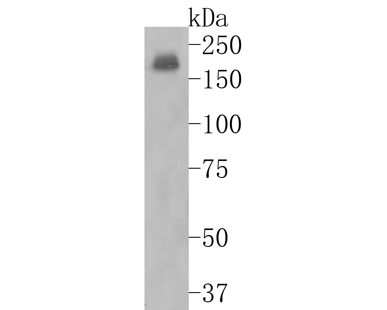
Western blot analysis of NMDAR2A on mouse hippocampus tissue lysates. Proteins were transferred to a PVDF membrane and blocked with 5% BSA in PBS for 1 hour at room temperature. The primary antibody (1/2000) was used in 5% BSA at room temperature for 2 hours. Goat Anti-Rabbit IgG - HRP Secondary Antibody at 1:50000 dilution was used for 1 hour at room temperature.IHC
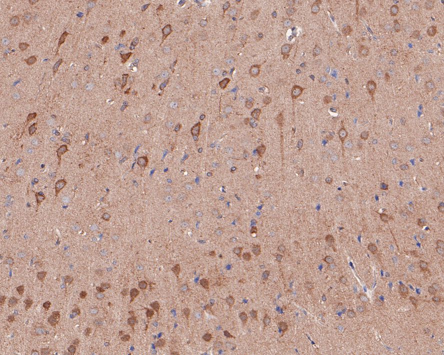
Immunohistochemical analysis of paraffin-embedded rat brain tissue with Rabbit anti-NMDAR2A antibody at 1/50 dilution. The section was pre-treated using heat mediated antigen retrieval with Tris-EDTA buffer (pH 9.0) for 20 minutes. The tissues were blocked in 1% BSA for 20 minutes at room temperature, washed with ddH2O and PBS, and then probed with the primary antibody at 1/50 dilution for 1 hour at room temperature. The detection was performed using an HRP conjugated compact polymer system. DAB was used as the chromogen. Tissues were counterstained with hematoxylin and mounted with DPX.IHC
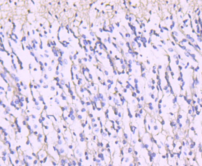
Immunohistochemical analysis of paraffin-embedded human brain tissue using anti-NMDAR2A antibody. Counter stained with hematoxylin.IHC
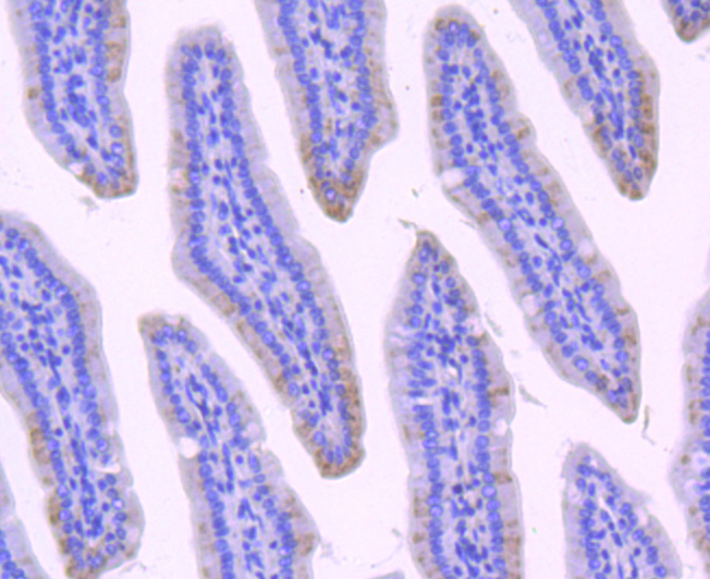
Immunohistochemical analysis of paraffin-embedded mouse small intestine tissue using anti-NMDAR2A antibody. Counter stained with hematoxylin.IF-P
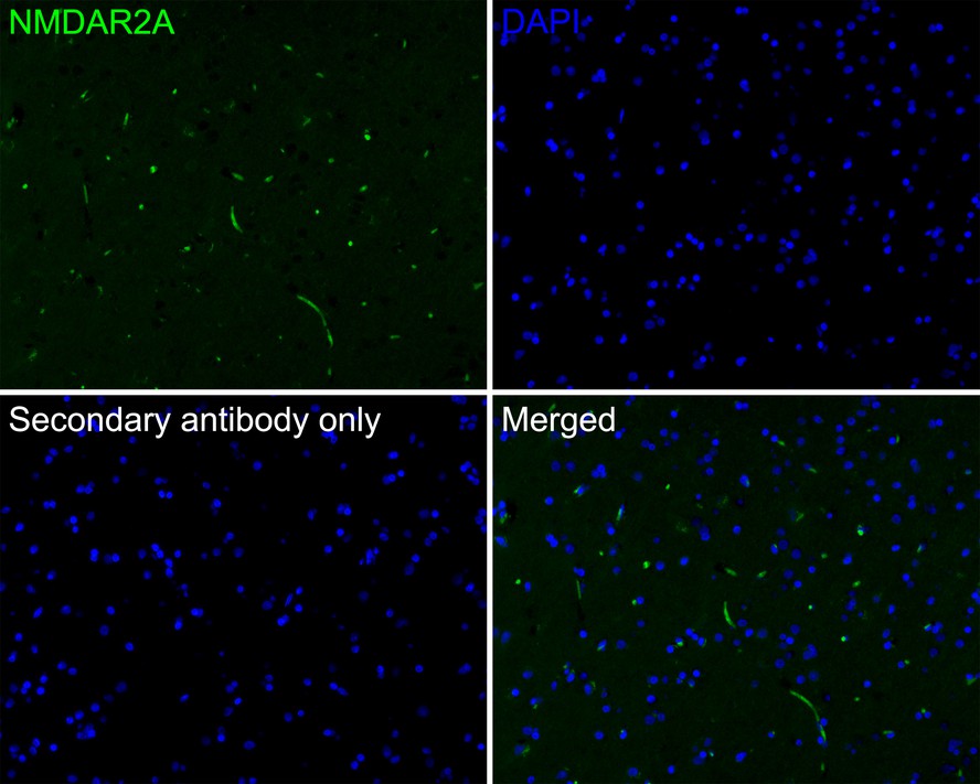
Immunofluorescence analysis of paraffin-embedded human brain tissue labeling NMDAR2A with Rabbit anti-NMDAR2A antibody at 1/200 dilution. The section was pre-treated using heat mediated antigen retrieval with Tris-EDTA buffer (pH 9.0) for 20 minutes. The tissues were blocked in 10% negative goat serum for 1 hour at room temperature, washed with PBS, and then probed with the primary antibody (green) at 1/200 dilution overnight at 4 ℃, washed with PBS. Goat Anti-Rabbit IgG H&L (488) was used as the secondary antibody at 1/1,000 dilution. Nuclei were counterstained with DAPI (blue).| Product Name | NMDAR2A Recombinant Rabbit Monoclonal Antibody |
|---|---|
| Antibody Type | Primary Antibodies |
| Immunogen | Synthetic peptide within Human NMDAR2A aa 1356-1405 / 1464. |
| Clonality | Monoclonal |
|---|---|
| Isotype | IgG |
| Host Species | Rabbit |
| Tested Applications | IF-PIHCWB |
| WB:1:500 IF-P:1:200 IHC:1:50-1:100 |
|
| Species Reactivity | HumanMouseRat |
| Concentration | 1mg/ml |
| Purification | Affinity purified |
| Gene Symbol | GRIN2A |
|---|---|
| Gene Synonyms | LKS EPND FESD NR2A GluN2A NMDAR2A |
| Gene Full Name | glutamate ionotropic receptor NMDA type subunit 2A |
| Gene Summary | This gene encodes a member of the glutamate-gated ion channel protein family. The encoded protein is an N-methyl-D-aspartate (NMDA) receptor subunit. NMDA receptors are both ligand-gated and voltage-dependent, and are involved in long-term potentiation, an activity-dependent increase in the efficiency of synaptic transmission thought to underlie certain kinds of memory and learning. These receptors are permeable to calcium ions, and activation results in a calcium influx into post-synaptic cells, which results in the activation of several signaling cascades. Disruption of this gene is associated with focal epilepsy and speech disorder with or without cognitive disability. Alternative splicing results in multiple transcript variants. [provided by RefSeq, May 2014] |
| Alternative Names | EPND antibody FESD antibody GluN2A antibody Glutamate [NMDA] receptor subunit epsilon-1 antibody Glutamate receptor antibody Glutamate receptor ionotropic N methyl D aspartate 2A antibody GRIN 2A antibody GRIN2A antibody hNR2A antibody LKS antibody EPND antibody FESD antibody GluN2A antibody Glutamate [NMDA] receptor subunit epsilon-1 antibody Glutamate receptor antibody Glutamate receptor ionotropic N methyl D aspartate 2A antibody GRIN 2A antibody GRIN2A antibody hNR2A antibody LKS antibody N methyl D aspartate receptor channel subunit epsilon 1 antibody N Methyl D Aspartate Receptor Subtype 2A antibody N methyl D aspartate receptor subunit 2A antibody N-methyl D-aspartate receptor subtype 2A antibody NMDA receptor subtype 2A antibody NMDAR 2A antibody NMDAR2A antibody NMDE1_HUMAN antibody NR2A antibody OTTHUMP00000160135 antibody OTTHUMP00000174531 antibody |
| Molecular Weight(MW) | 165kDa |
| Cellular Localization | Cell membrane, postsynaptic cell membrane, dendritic spine, synapse, Cytoplasmic vesicle membrane. |
WB
Western blot analysis of NMDAR2A on mouse hippocampus tissue lysates. Proteins were transferred to a PVDF membrane and blocked with 5% BSA in PBS for 1 hour at room temperature. The primary antibody (1/2000) was used in 5% BSA at room temperature for 2 hours. Goat Anti-Rabbit IgG - HRP Secondary Antibody at 1:50000 dilution was used for 1 hour at room temperature.
IHC
Immunohistochemical analysis of paraffin-embedded rat brain tissue with Rabbit anti-NMDAR2A antibody at 1/50 dilution. The section was pre-treated using heat mediated antigen retrieval with Tris-EDTA buffer (pH 9.0) for 20 minutes. The tissues were blocked in 1% BSA for 20 minutes at room temperature, washed with ddH2O and PBS, and then probed with the primary antibody at 1/50 dilution for 1 hour at room temperature. The detection was performed using an HRP conjugated compact polymer system. DAB was used as the chromogen. Tissues were counterstained with hematoxylin and mounted with DPX.
IHC
Immunohistochemical analysis of paraffin-embedded human brain tissue using anti-NMDAR2A antibody. Counter stained with hematoxylin.
IHC
Immunohistochemical analysis of paraffin-embedded mouse small intestine tissue using anti-NMDAR2A antibody. Counter stained with hematoxylin.
IF-P
Immunofluorescence analysis of paraffin-embedded human brain tissue labeling NMDAR2A with Rabbit anti-NMDAR2A antibody at 1/200 dilution. The section was pre-treated using heat mediated antigen retrieval with Tris-EDTA buffer (pH 9.0) for 20 minutes. The tissues were blocked in 10% negative goat serum for 1 hour at room temperature, washed with PBS, and then probed with the primary antibody (green) at 1/200 dilution overnight at 4 ℃, washed with PBS. Goat Anti-Rabbit IgG H&L (488) was used as the secondary antibody at 1/1,000 dilution. Nuclei were counterstained with DAPI (blue).| Application Notes | WB:1:500 IF-P:1:200 IHC:1:50-1:100 |
|---|
| Form | Liquid |
|---|---|
| Storage Instructions | Shipped at 4℃. Store at +4℃ short term (1-2 weeks). It is recommended to aliquot into single-use upon delivery. Store at -20℃ long term. |
| Storage Buffer | 1*TBS (pH7.4), 0.05% BSA, 40% Glycerol. Preservative: 0.05% Sodium Azide. |


