WB
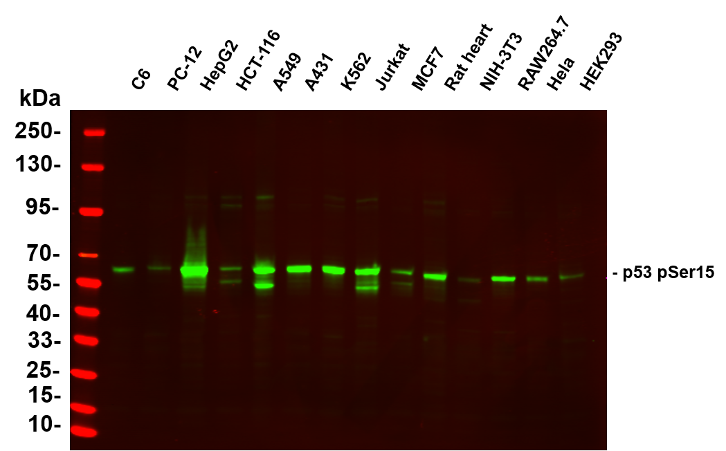
Various whole cell lysates were separated by 4-20% SDS-PAGE, and the primary antibody was used at 4~C, over night with a 1:5000 dilution . The Dylight 800-conjugated Goat anti-Rabbit antibod was used to detect the antibody. Lane1:C6- Rat glioma cells, Lane2: PC-12 - Pheochromocytoma in rats, Lane3: HepG2 - Human hepatocellular carcinoma,Lane4: HCT-116 - Human colon cancer,Lane5: A549 - Human lung adenocarcinoma,Lane6: A431 - Human skin squamous cell carcinoma,Lane7: K562 - Human chronic myeloid leukemia,Lane8: Jurkat - Human acute T cell leukemia cells,Lane9: MCF7 - Human breast cancer,Lane10: Rat heart - Human neuroblastoma cells,Lane11: NIH-3T3 - NIH mouse fibroblasts,Lane12: RAW264.7 - Mouse mononuclear macrophage leukemia cells,Lane13: Hela - Human cervical cancer Lane,14: HEK293 - Human normal embryonic kidney.WB
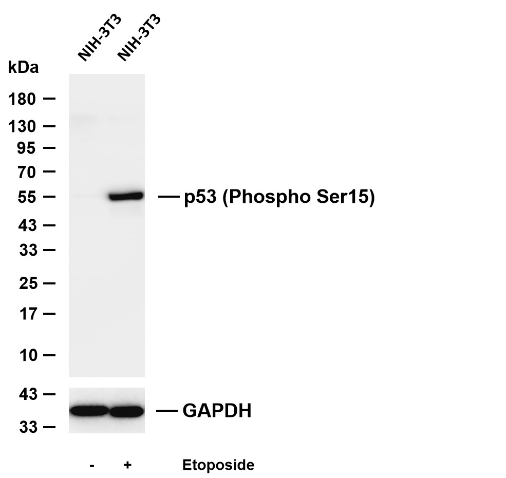
Various whole cell lysates were separated by 4-20% SDS-PAGE, and the membrane was blotted with anti-p53 (Phospho Ser15) antibody. The HRP-conjugated Goat anti-Rabbit IgG (H + L) antibody was used to detect the antibody. Lane 1: NIH-3T3 Lane 2: NIH-3T3 was treated with Etoposide(25µM) for 3 hours.ICC/IF
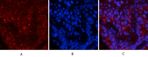
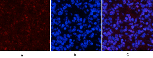
Immunofluorescence analysis of rat-lung tissue. 1,p53 (phospho Ser15) Antibody(red) was diluted at 1:200(4℃,overnight). 2, Cy3 labled Secondary antibody was diluted at 1:300(room temperature, 50min).3, Picture B: DAPI(blue) 10min. Picture A:Target. Picture B: DAPI. Picture C: merge of A+B.ICC/IF
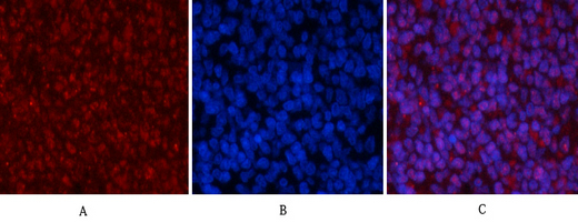
Immunofluorescence analysis of rat-spleen tissue. 1,p53 (phospho Ser15) Antibody(red) was diluted at 1:200(4℃,overnight). 2, Cy3 labled Secondary antibody was diluted at 1:300(room temperature, 50min).3, Picture B: DAPI(blue) 10min. Picture A:Target. Picture B: DAPI. Picture C: merge of A+B.ICC/IF
Immunofluorescence analysis of mouse-spleen tissue. 1,p53 (phospho Ser15) Antibody(red) was diluted at 1:200(4℃,overnight). 2, Cy3 labled Secondary antibody was diluted at 1:300(room temperature, 50min).3, Picture B: DAPI(blue) 10min. Picture A:Target. Picture B: DAPI. Picture C: merge of A+B.IHC
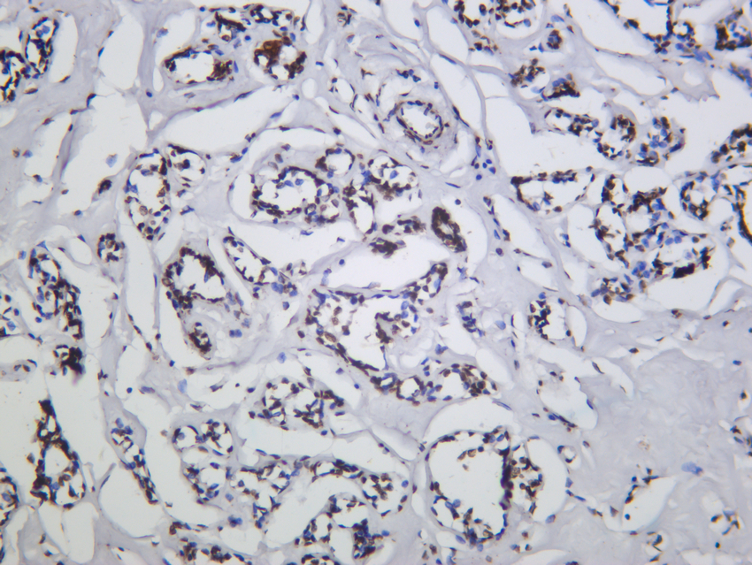
Human breast carcinoma was stained with anti-p53 (Phospho Ser15) Rabbit antibody.| Product Name | p53 (Phospho Ser15) Rabbit mAb |
|---|---|
| Antibody Type | Primary Antibodies |
| Clonality | monoclonal |
|---|---|
| Isotype | IgG,K |
| Host Species | Rabbit |
| Tested Applications | ICC/IFIHCWB |
| WB:1:2000-1:10000 IHC:1:200-1:1000 ICC/IF:1:200-1:1000 |
|
| Species Reactivity | HumanMouseRat |
| Concentration | 1mg/ml |
| Purification | Protein A |
| Gene Symbol | TP53 |
|---|---|
| Gene Synonyms | P53 BCC7 LFS1 BMFS5 TRP53 |
| Gene Full Name | tumor protein p53 |
| Gene Summary | This gene encodes a tumor suppressor protein containing transcriptional activation, DNA binding, and oligomerization domains. The encoded protein responds to diverse cellular stresses to regulate expression of target genes, thereby inducing cell cycle arrest, apoptosis, senescence, DNA repair, or changes in metabolism. Mutations in this gene are associated with a variety of human cancers, including hereditary cancers such as Li-Fraumeni syndrome. Alternative splicing of this gene and the use of alternate promoters result in multiple transcript variants and isoforms. Additional isoforms have also been shown to result from the use of alternate translation initiation codons from identical transcript variants (PMIDs: 12032546, 20937277). [provided by RefSeq, Dec 2016] |
| Alternative Names | Antigen NY-CO-13 BCC7 Cellular tumor antigen p53 FLJ92943 LFS1 Mutant tumor protein 53 p53 p53 tumor suppressor P53_HUMAN Phosphoprotein p53 Tp53 Transformation related protein 53 TRP53 tumor antigen p55 Tumor protein 53 Tumor protein p53 Tumor suppressor p53 |
| Molecular Weight(MW) | 53kDa(Observed MW 55kDa) |
| Cellular Localization | Nuclear. |
WB
Various whole cell lysates were separated by 4-20% SDS-PAGE, and the primary antibody was used at 4~C, over night with a 1:5000 dilution . The Dylight 800-conjugated Goat anti-Rabbit antibod was used to detect the antibody. Lane1:C6- Rat glioma cells, Lane2: PC-12 - Pheochromocytoma in rats, Lane3: HepG2 - Human hepatocellular carcinoma,Lane4: HCT-116 - Human colon cancer,Lane5: A549 - Human lung adenocarcinoma,Lane6: A431 - Human skin squamous cell carcinoma,Lane7: K562 - Human chronic myeloid leukemia,Lane8: Jurkat - Human acute T cell leukemia cells,Lane9: MCF7 - Human breast cancer,Lane10: Rat heart - Human neuroblastoma cells,Lane11: NIH-3T3 - NIH mouse fibroblasts,Lane12: RAW264.7 - Mouse mononuclear macrophage leukemia cells,Lane13: Hela - Human cervical cancer Lane,14: HEK293 - Human normal embryonic kidney.
WB
Various whole cell lysates were separated by 4-20% SDS-PAGE, and the membrane was blotted with anti-p53 (Phospho Ser15) antibody. The HRP-conjugated Goat anti-Rabbit IgG (H + L) antibody was used to detect the antibody. Lane 1: NIH-3T3 Lane 2: NIH-3T3 was treated with Etoposide(25µM) for 3 hours.
ICC/IF
Immunofluorescence analysis of rat-lung tissue. 1,p53 (phospho Ser15) Antibody(red) was diluted at 1:200(4℃,overnight). 2, Cy3 labled Secondary antibody was diluted at 1:300(room temperature, 50min).3, Picture B: DAPI(blue) 10min. Picture A:Target. Picture B: DAPI. Picture C: merge of A+B.
ICC/IF
Immunofluorescence analysis of rat-spleen tissue. 1,p53 (phospho Ser15) Antibody(red) was diluted at 1:200(4℃,overnight). 2, Cy3 labled Secondary antibody was diluted at 1:300(room temperature, 50min).3, Picture B: DAPI(blue) 10min. Picture A:Target. Picture B: DAPI. Picture C: merge of A+B.
ICC/IF
Immunofluorescence analysis of mouse-spleen tissue. 1,p53 (phospho Ser15) Antibody(red) was diluted at 1:200(4℃,overnight). 2, Cy3 labled Secondary antibody was diluted at 1:300(room temperature, 50min).3, Picture B: DAPI(blue) 10min. Picture A:Target. Picture B: DAPI. Picture C: merge of A+B.
IHC
Human breast carcinoma was stained with anti-p53 (Phospho Ser15) Rabbit antibody.| Application Notes | WB:1:2000-1:10000 IHC:1:200-1:1000 ICC/IF:1:200-1:1000 |
|---|
| Form | Liquid |
|---|---|
| Storage Instructions | -15°C to -25°C/1 year(Do not lower than -25°C) |
| Storage Buffer | PBS, 50% glycerol, 0.05% Proclin 300, 0.05%BSA |


