WB
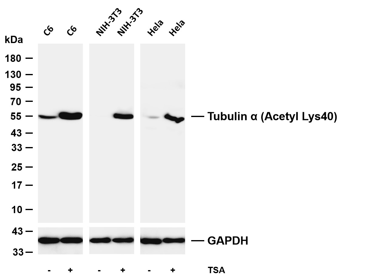
Various whole cell lysates were separated by 4-20% SDS-PAGE, and the membrane was blotted with anti-Tubulin α (Acetyl Lys40) antibody. The HRP-conjugated Goat anti-Rabbit IgG (H + L) antibody was used to detect the antibody. Lane 1: C6 Lane 2: C6 was treated with TSA (500ng/mL) for 4 hours Lane 3: NIH-3T3 Lane 4: NIH-3T3 was treated with TSA (400nM) for 18 hours Lane 5: Hela Lane 6: Hela was treated with TSA (400nM) for 18 hours.IHC
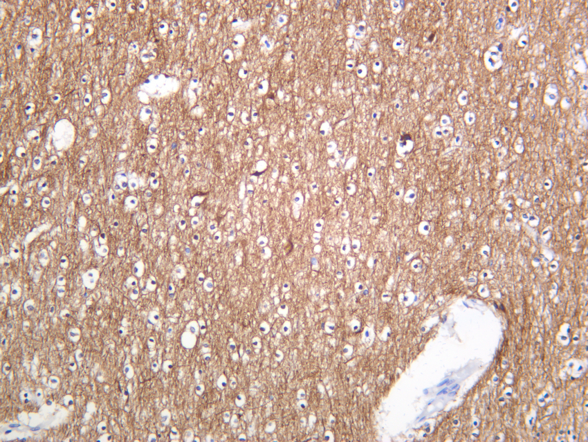
Human brain was stained with anti-Tubulin α (Acetyl Lys40) Rabbit antibody.ICC/IF
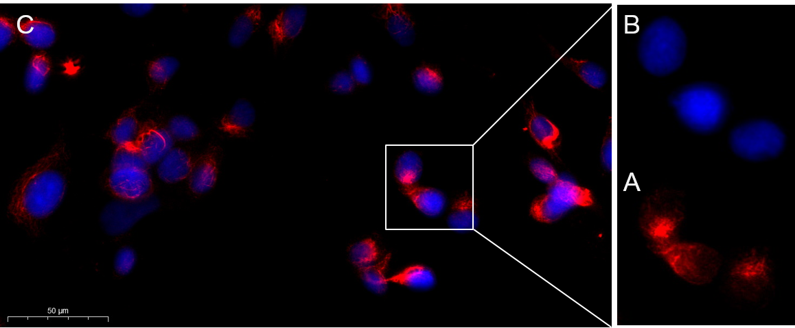
Immunofluorescence analysis of MCF7 . Picture A: Tubulin α (Acetyl Lys40) Rabbit mAb (red). Picture B: DAPI (blue). Picture C: Merge of A+B.| Product Name | α Tubulin (Acetyl Lys40) Rabbit mAb |
|---|---|
| Antibody Type | Primary Antibodies |
| Clonality | monoclonal |
|---|---|
| Isotype | IgG,K |
| Host Species | Rabbit |
| Tested Applications | ICC/IFIHCWB |
| WB:1:2000-1:10000 ICC/IF:1:200-1:1000 IHC:1:200-1:1000 |
|
| Species Reactivity | HumanMouseRat |
| Concentration | 1mg/ml |
| Purification | Protein A |
| Gene Symbol | TUBA4A |
|---|---|
| Gene Synonyms | ALS22 TUBA1 CMYO26 SPAX11 FTDALS9 OZEMA23 H2-ALPHA |
| Gene Full Name | tubulin alpha 4a |
| Gene Summary | Microtubules of the eukaryotic cytoskeleton perform essential and diverse functions and are composed of a heterodimer of alpha and beta tubulin. The genes encoding these microtubule constituents are part of the tubulin superfamily, which is composed of six distinct families. Genes from the alpha, beta and gamma tubulin families are found in all eukaryotes. The alpha and beta tubulins represent the major components of microtubules, while gamma tubulin plays a critical role in the nucleation of microtubule assembly. There are multiple alpha and beta tubulin genes and they are highly conserved among and between species. This gene encodes an alpha tubulin that is a highly conserved homolog of a rat testis-specific alpha tubulin. Alternatively spliced transcript variants encoding different isoforms have been found for this gene. [provided by RefSeq, Jun 2013] |
| Alternative Names | Alpha Tubulin |
| Molecular Weight(MW) | 50kDa(Observed MW 55kDa) |
| Cellular Localization | Cytoplasm |
WB
Various whole cell lysates were separated by 4-20% SDS-PAGE, and the membrane was blotted with anti-Tubulin α (Acetyl Lys40) antibody. The HRP-conjugated Goat anti-Rabbit IgG (H + L) antibody was used to detect the antibody. Lane 1: C6 Lane 2: C6 was treated with TSA (500ng/mL) for 4 hours Lane 3: NIH-3T3 Lane 4: NIH-3T3 was treated with TSA (400nM) for 18 hours Lane 5: Hela Lane 6: Hela was treated with TSA (400nM) for 18 hours.
IHC
Human brain was stained with anti-Tubulin α (Acetyl Lys40) Rabbit antibody.
ICC/IF
Immunofluorescence analysis of MCF7 . Picture A: Tubulin α (Acetyl Lys40) Rabbit mAb (red). Picture B: DAPI (blue). Picture C: Merge of A+B.| Application Notes | WB:1:2000-1:10000 ICC/IF:1:200-1:1000 IHC:1:200-1:1000 |
|---|
| Form | Liquid |
|---|---|
| Storage Instructions | -15°C to -25°C/1 year(Do not lower than -25°C) |
| Storage Buffer | PBS, 50% glycerol, 0.05% Proclin 300, 0.05%BSA |


