WB
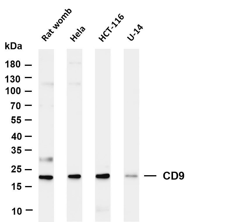
Various whole cell lysates were separated by 4-20% SDS-PAGE, and the membrane was blotted with anti-CD9 antibody. The HRP-conjugated Goat anti-Rabbit IgG(H + L) antibody was used to detect the antibody. Lane 1: Rat womb Lane 2: Hela Lane 3: HCT-116 Lane 4: U-14.ICC/IF
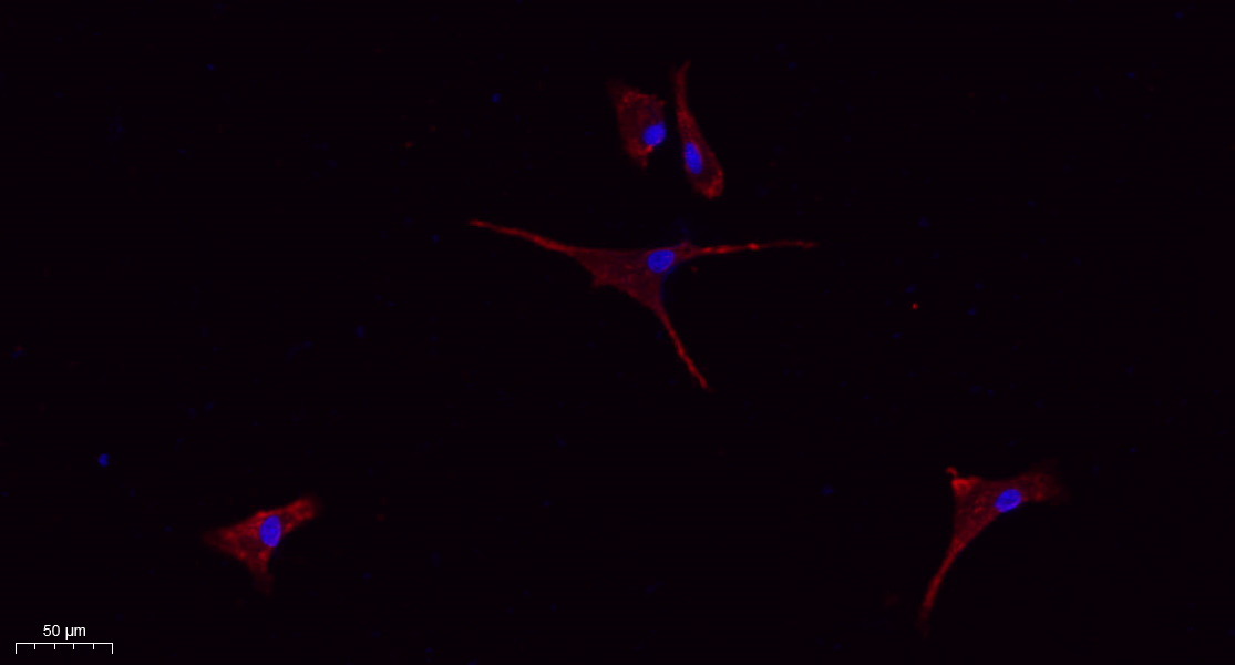
Immunofluorescence analysis of A549. 1,primary Antibody(red) was diluted at 1:200(4℃ overnight). 2, Goat Anti Rabbit IgG (H&L) - Alexa Fluor 594 Secondary antibody was diluted at 1:1000(room temperature, 50min).3, DAPI(blue) 10min.ICC/IF
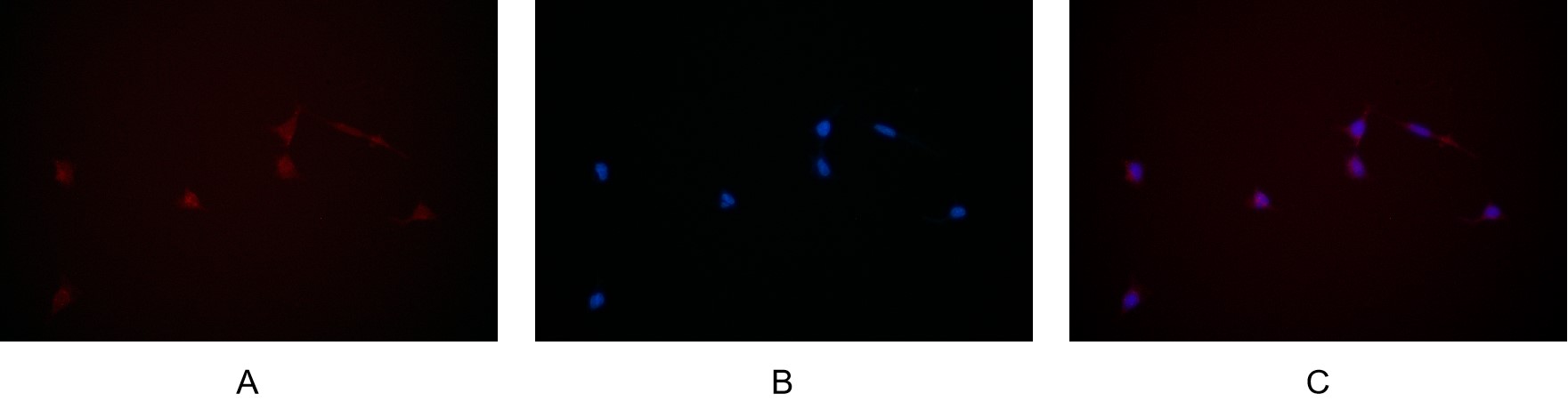
Immunofluorescence analysis of HEK293. Picture A: CD9 antibody (red). Picture B: DAPI (blue). Picture C: Merge of A+B.IHC
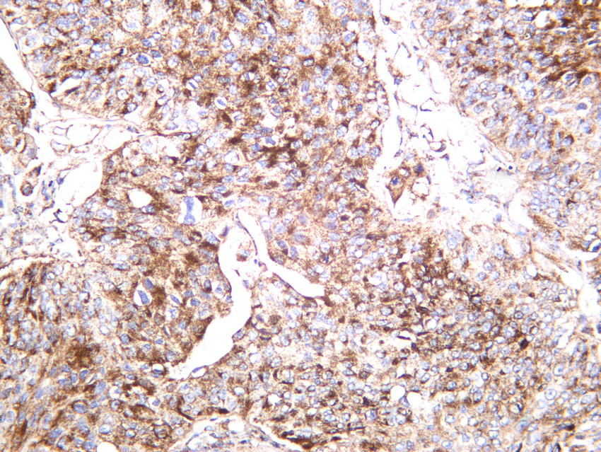
Human bladder carcinoma was stained with anti-CD9 rabbit antibody.| Product Name | CD9 Rabbit mAb |
|---|---|
| Antibody Type | Primary Antibodies |
| Clonality | monoclonal |
|---|---|
| Isotype | IgG,K |
| Host Species | Rabbit |
| Tested Applications | ICC/IFIHCWB |
| WB:1:2000-1:10000 IHC:1:200-1:1000 ICC/IF:1:200-1:1000 |
|
| Species Reactivity | BovineHumanMouseRat |
| Concentration | 1mg/ml |
| Purification | Protein A |
| Gene Symbol | CD9 |
|---|---|
| Gene Synonyms | MIC3 MRP-1 BTCC-1 DRAP-27 TSPAN29 TSPAN-29 |
| Gene Full Name | CD9 molecule |
| Gene Summary | This gene encodes a member of the transmembrane 4 superfamily, also known as the tetraspanin family. Tetraspanins are cell surface glycoproteins with four transmembrane domains that form multimeric complexes with other cell surface proteins. The encoded protein functions in many cellular processes including differentiation, adhesion, and signal transduction, and expression of this gene plays a critical role in the suppression of cancer cell motility and metastasis. [provided by RefSeq, Jan 2011] |
| Alternative Names | CD9 MIC3 TSPAN29 GIG2 CD9 antigen 5H9 antigen Cell growth-inhibiting gene 2 protein Leukocyte antigen MIC3 Motility-related protein MRP-1 Tetraspanin-29 Tspan-29 p24 CD antigen CD9 |
| Molecular Weight(MW) | 25kDa(Observed MW 20kDa) |
| Cellular Localization | Membrane. |
WB
Various whole cell lysates were separated by 4-20% SDS-PAGE, and the membrane was blotted with anti-CD9 antibody. The HRP-conjugated Goat anti-Rabbit IgG(H + L) antibody was used to detect the antibody. Lane 1: Rat womb Lane 2: Hela Lane 3: HCT-116 Lane 4: U-14.
ICC/IF
Immunofluorescence analysis of A549. 1,primary Antibody(red) was diluted at 1:200(4℃ overnight). 2, Goat Anti Rabbit IgG (H&L) - Alexa Fluor 594 Secondary antibody was diluted at 1:1000(room temperature, 50min).3, DAPI(blue) 10min.
ICC/IF
Immunofluorescence analysis of HEK293. Picture A: CD9 antibody (red). Picture B: DAPI (blue). Picture C: Merge of A+B.
IHC
Human bladder carcinoma was stained with anti-CD9 rabbit antibody.| Application Notes | WB:1:2000-1:10000 IHC:1:200-1:1000 ICC/IF:1:200-1:1000 |
|---|
| Form | Liquid |
|---|---|
| Storage Instructions | -15°C to -25°C/1 year(Do not lower than -25°C) |
| Storage Buffer | PBS, 50% glycerol, 0.05% Proclin 300, 0.05%BSA |


