WB
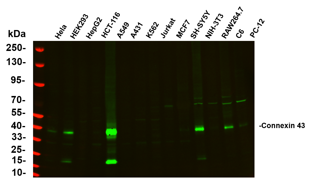
Various whole cell lysates were separated by 4-20% SDS-PAGE, and the primary antibody was used at 4℃, over night with a 1:5000 dilution . The Dylight 800-conjugated Goat anti-Rabbit antibody was used to detect the antibody. Lane1: Hela - Human cervical cancer Lane2: HEK293 - Human normal embryonic kidney Lane3: HepG2 - Human hepatocellular carcinoma Lane4: HCT-116 - Human colon cancer Lane5: A549 - Human lung adenocarcinoma Lane6: A431 - Human skin squamous cell carcinoma Lane7: K562 - Human chronic myeloid leukemia Lane8: Jurkat - Human acute T cell leukemia cells Lane9: MCF7 - Human breast cancer Lane10: SH-SY5Y - Human neuroblastoma cells Lane11: NIH-3T3 - NIH mouse fibroblasts Lane12: RAW264.7 - Mouse mononuclear macrophage leukemia cells Lane13: C6 - Rat glioma cells Lane14: PC-12 - Pheochromocytoma in rats.WB
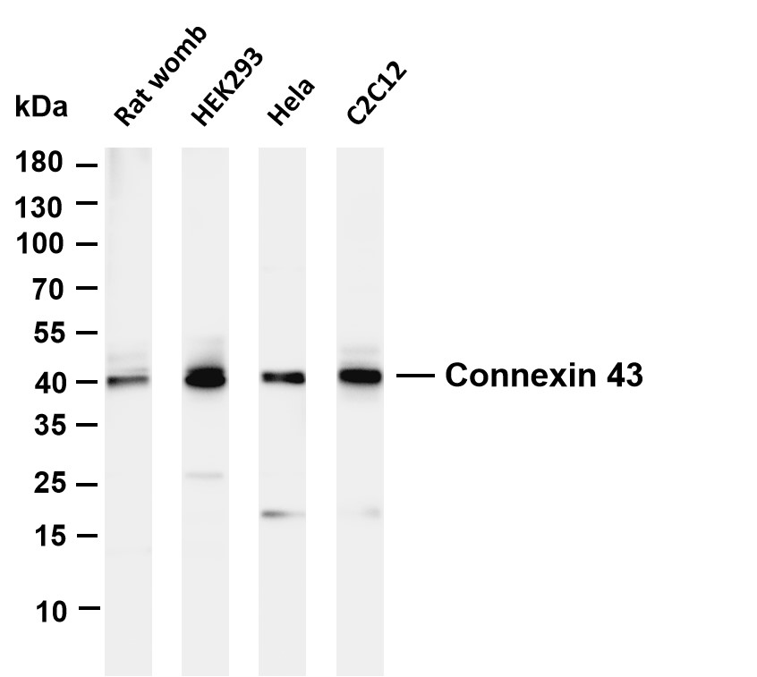
Various whole cell lysates were separated by 4-20% SDS-PAGE, and the membrane was blotted with anti-Connexin 43 antibody. The HRP-conjugated Goat anti-Rabbit IgG(H + L) antibody was used to detect the antibody. Lane 1: Rat womb Lane 2: HEK293 Lane 3: Hela Lane 4: C2C12.IHC
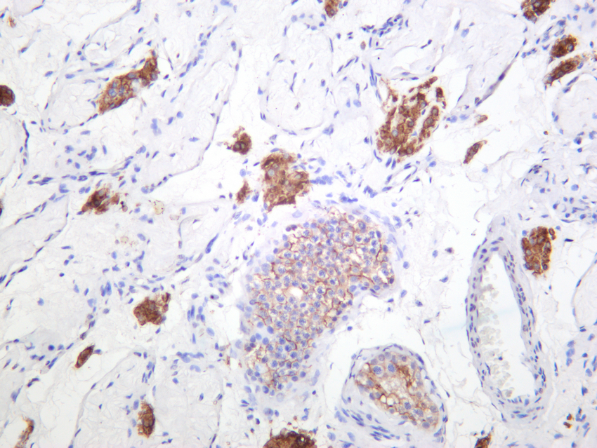
Human testis was stained with anti-Connexin 43 rabbit antibody.IHC
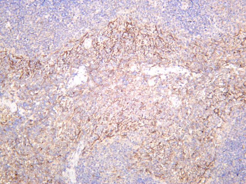
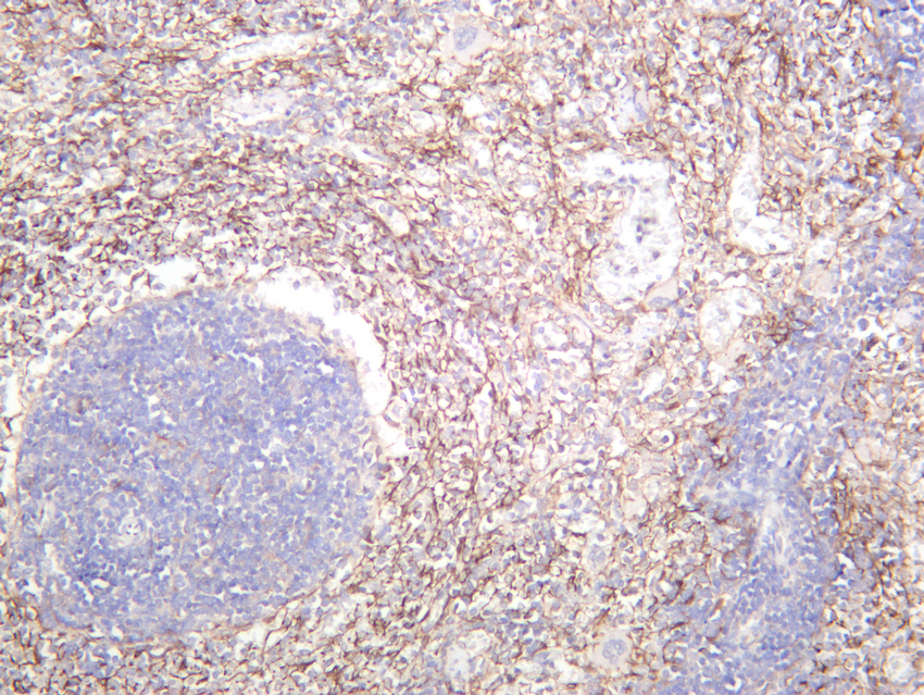
Mouse spleen was stained with anti-Connexin 43 rabbit antibody.IHC
Rat spleen was stained with anti-Connexin 43 rabbit antibody.ICC/IF
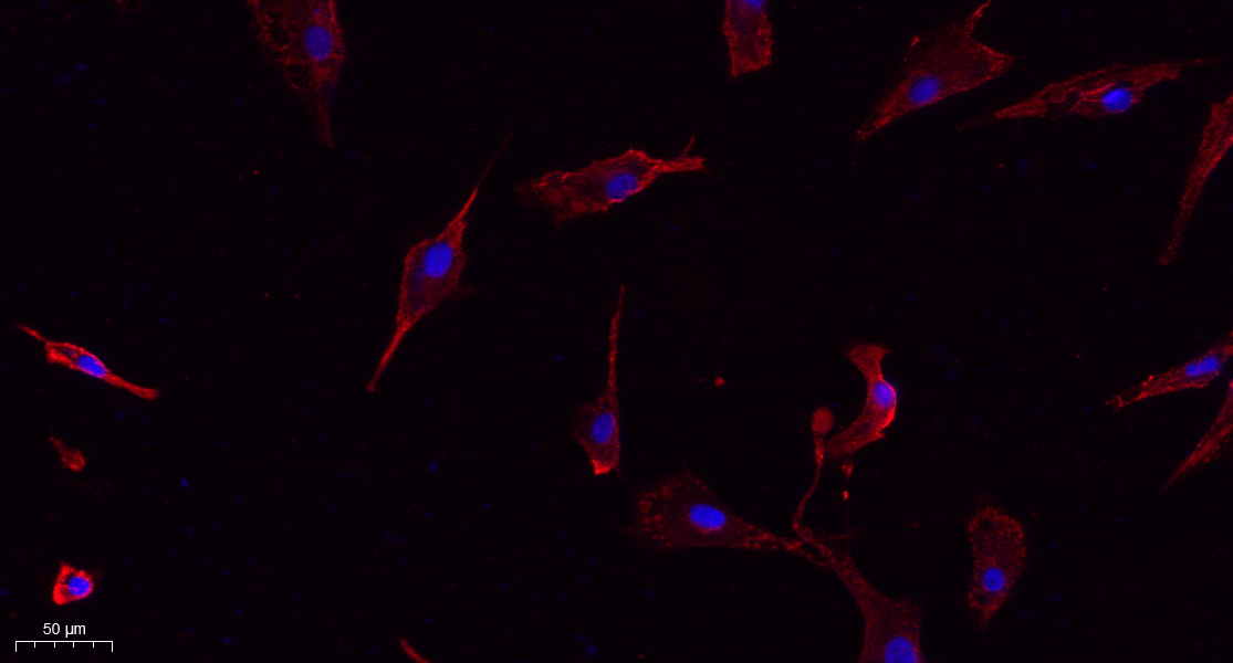
Immunofluorescence analysis of A549. 1,primary Antibody(red) was diluted at 1:200(4℃ overnight). 2, Goat Anti Rabbit IgG (H&L) - Alexa Fluor 594 Secondary antibody was diluted at 1:1000(room temperature, 50min).3, DAPI(blue) 10min.ICC/IF
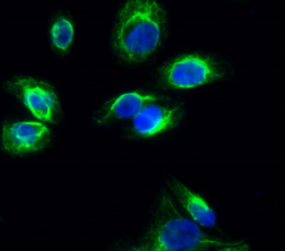
Immunofluorescence analysis of Hela cell. 1,Connexin 43 Antibody(green) was diluted at 1:200(4° overnight). 2, Goat Anti Rabbit Alexa Fluor 488 was diluted at 1:1000(room temperature, 50min). 3 DAPI(blue) 10min.ICC/IF
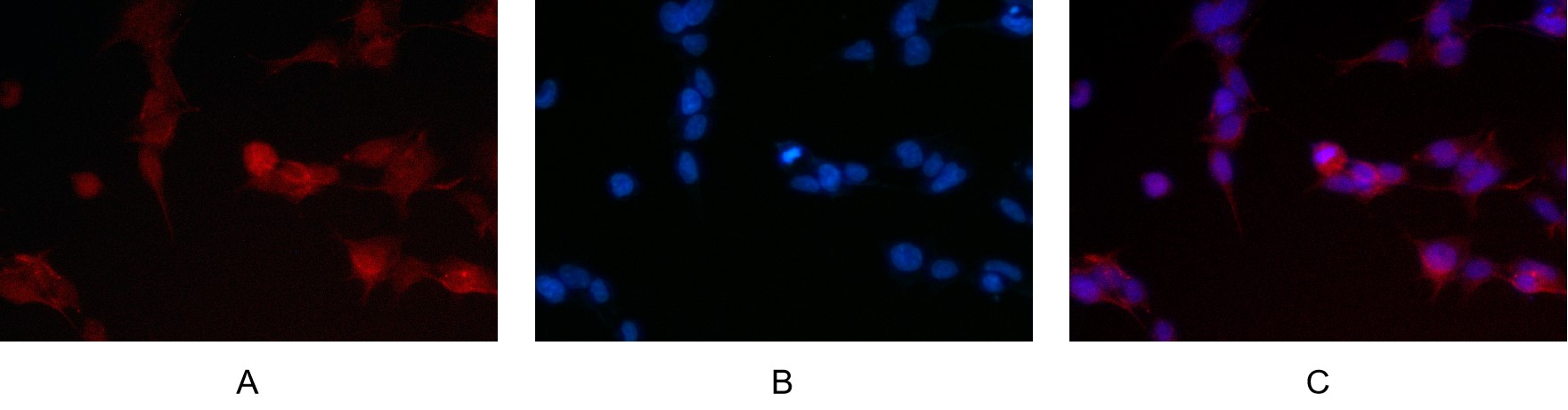
Immunofluorescence analysis of HEK293. Picture A: Connexin 43 antibody (red). Picture B: DAPI (blue). Picture C: Merge of A+B.| Product Name | Connexin 43 Rabbit mAb |
|---|---|
| Antibody Type | Primary Antibodies |
| Clonality | monoclonal |
|---|---|
| Isotype | IgG,K |
| Host Species | Rabbit |
| Tested Applications | ICC/IFIHCWB |
| WB:1:2000-1:10000 IHC:1:200-1000 ICC/IF:1:200-1000 |
|
| Species Reactivity | HumanMouseRat |
| Concentration | 1mg/ml |
| Purification | Protein A |
| Gene Symbol | GJA1 |
|---|---|
| Gene Synonyms | HSS CMDR CX43 EKVP GJAL ODDD AVSD3 EKVP3 HLHS1 PPKCA |
| Gene Full Name | gap junction protein alpha 1 |
| Gene Summary | This gene is a member of the connexin gene family. The encoded protein is a component of gap junctions, which are composed of arrays of intercellular channels that provide a route for the diffusion of low molecular weight materials from cell to cell. The encoded protein is the major protein of gap junctions in the heart that are thought to have a crucial role in the synchronized contraction of the heart and in embryonic development. A related intronless pseudogene has been mapped to chromosome 5. Mutations in this gene have been associated with oculodentodigital dysplasia, autosomal recessive craniometaphyseal dysplasia and heart malformations. [provided by RefSeq, May 2014] |
| Alternative Names | GJA1 GJAL Gap junction alpha-1 protein Connexin-43 Cx43 Gap junction 43 kDa heart protein |
| Molecular Weight(MW) | 43kDa(Observed MW 43kDa) |
| Cellular Localization | Membrane |
WB
Various whole cell lysates were separated by 4-20% SDS-PAGE, and the primary antibody was used at 4℃, over night with a 1:5000 dilution . The Dylight 800-conjugated Goat anti-Rabbit antibody was used to detect the antibody. Lane1: Hela - Human cervical cancer Lane2: HEK293 - Human normal embryonic kidney Lane3: HepG2 - Human hepatocellular carcinoma Lane4: HCT-116 - Human colon cancer Lane5: A549 - Human lung adenocarcinoma Lane6: A431 - Human skin squamous cell carcinoma Lane7: K562 - Human chronic myeloid leukemia Lane8: Jurkat - Human acute T cell leukemia cells Lane9: MCF7 - Human breast cancer Lane10: SH-SY5Y - Human neuroblastoma cells Lane11: NIH-3T3 - NIH mouse fibroblasts Lane12: RAW264.7 - Mouse mononuclear macrophage leukemia cells Lane13: C6 - Rat glioma cells Lane14: PC-12 - Pheochromocytoma in rats.
WB
Various whole cell lysates were separated by 4-20% SDS-PAGE, and the membrane was blotted with anti-Connexin 43 antibody. The HRP-conjugated Goat anti-Rabbit IgG(H + L) antibody was used to detect the antibody. Lane 1: Rat womb Lane 2: HEK293 Lane 3: Hela Lane 4: C2C12.
IHC
Human testis was stained with anti-Connexin 43 rabbit antibody.
IHC
Mouse spleen was stained with anti-Connexin 43 rabbit antibody.
IHC
Rat spleen was stained with anti-Connexin 43 rabbit antibody.
ICC/IF
Immunofluorescence analysis of A549. 1,primary Antibody(red) was diluted at 1:200(4℃ overnight). 2, Goat Anti Rabbit IgG (H&L) - Alexa Fluor 594 Secondary antibody was diluted at 1:1000(room temperature, 50min).3, DAPI(blue) 10min.
ICC/IF
Immunofluorescence analysis of Hela cell. 1,Connexin 43 Antibody(green) was diluted at 1:200(4° overnight). 2, Goat Anti Rabbit Alexa Fluor 488 was diluted at 1:1000(room temperature, 50min). 3 DAPI(blue) 10min.
ICC/IF
Immunofluorescence analysis of HEK293. Picture A: Connexin 43 antibody (red). Picture B: DAPI (blue). Picture C: Merge of A+B.| Application Notes | WB:1:2000-1:10000 IHC:1:200-1000 ICC/IF:1:200-1000 |
|---|
| Form | Liquid |
|---|---|
| Storage Instructions | -15°C to -25°C/1 year(Do not lower than -25°C) |
| Storage Buffer | PBS, 50% glycerol, 0.05% Proclin 300, 0.05%BSA |


