WB
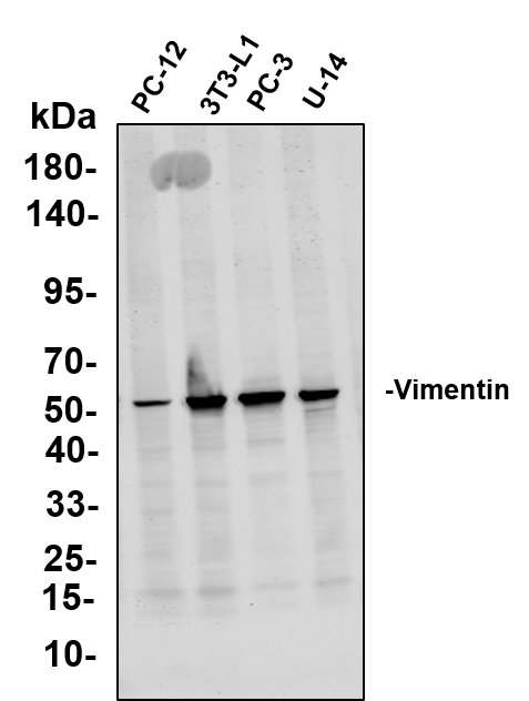
Various whole cell lysates were separated by 4-20% SDS-PAGE, and the primary antibody was used at 4℃, over night with a 1:5000 dilution . The Dylight 800-conjugated Goat anti-Rabbit antibody was used to detect the antibody. Lane1: PC-12 - Rat adrenal pheochromocytoma Lane2: 3T3-L1 - Mouse embryonic fibroblast Lane3: PC-3 - Human prostate adenocarcinoma Lane4: U-14 - Mouse cervical carcinoma.WB
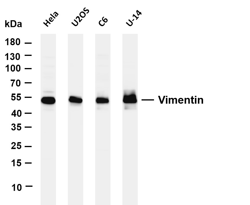
Various whole cell lysates were separated by 4-20% SDS-PAGE, and the membrane was blotted with anti-Vimentin antibody. The HRP-conjugated Goat anti-Rabbit IgG(H + L) antibody was used to detect the antibody. Lane 1: Hela Lane 2: U2OS Lane 3: C6 Lane 4: U-14.WB
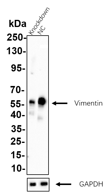
Western blot analysis of lysates from Hela WT and knockdown cell. Cell lysates were separated by 4-20% SDS-PAGE, and the membrane was blotted with Vimentin Rabbit mAb. The HRP-conjugated Goat anti-Rabbit IgG (H + L) antibody was used to detect the antibody.IHC
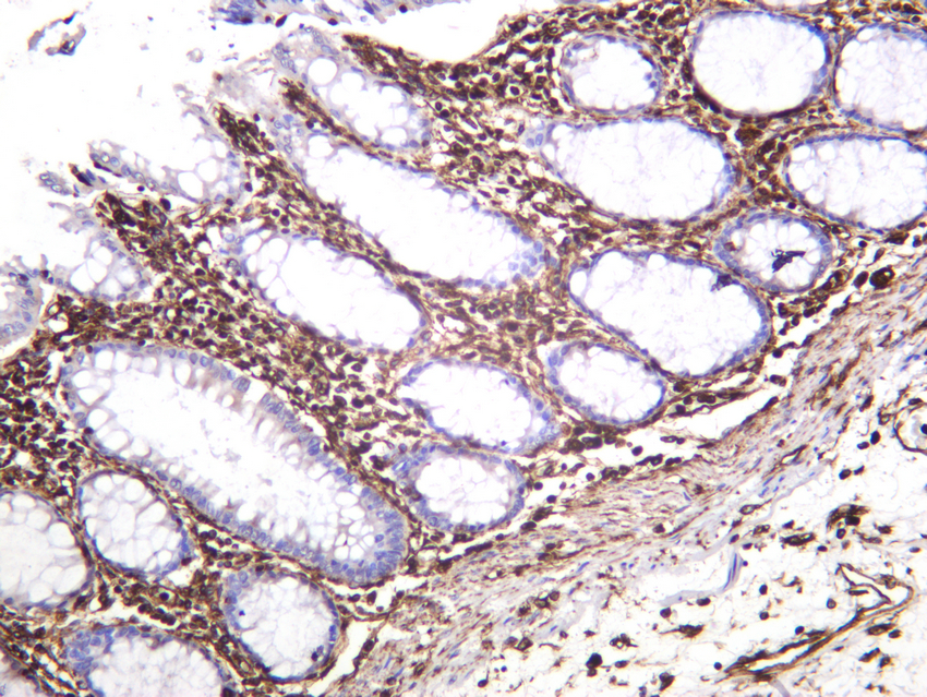
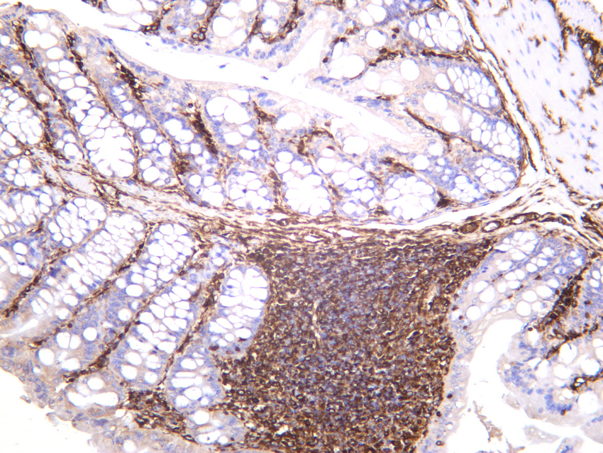
Human colon was stained with anti-Vimentin rabbit antibody.IHC
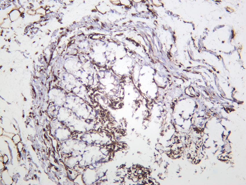
Mouse colon was stained with anti-Vimentin rabbit antibody.IHC
Rat colon was stained with anti-Vimentin rabbit antibody.ICC/IF
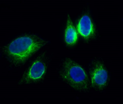
Immunofluorescence analysis of Hela cell. 1,Vimentin Antibody(green) was diluted at 1:200(4° overnight). 2, Goat Anti Rabbit Alexa Fluor 488 was diluted at 1:1000(room temperature, 50min). 3 DAPI(blue) 10min.ICC/IF
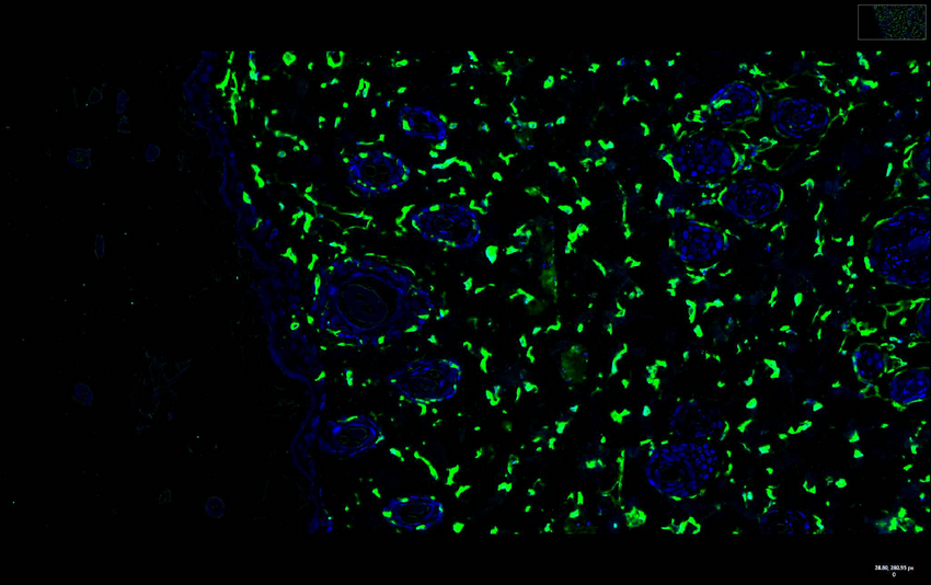
Rat skin was stained with anti-Vimentin Rabbit antibody.ICC/IF
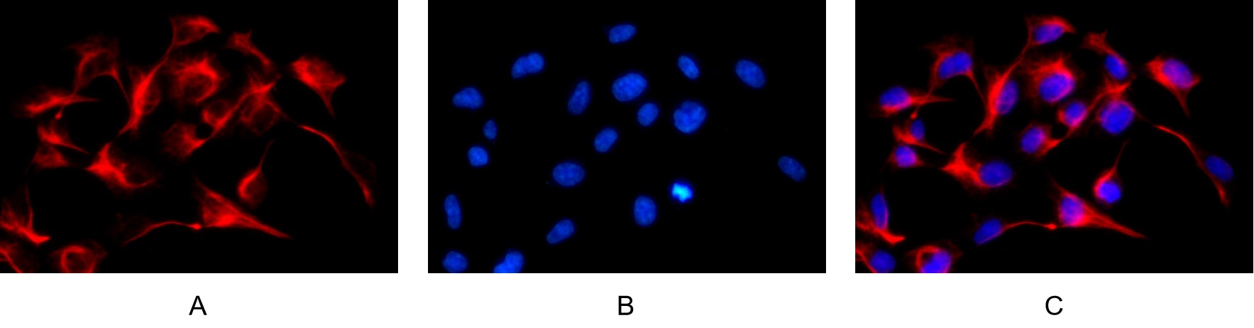
Immunofluorescence analysis of HEK293. Picture A: Vimentin antibody (red). Picture B: DAPI (blue). Picture C: Merge of A+B.| Product Name | Vimentin Rabbit mAb |
|---|---|
| Antibody Type | Primary Antibodies |
| Clonality | monoclonal |
|---|---|
| Isotype | IgG,K |
| Host Species | Rabbit |
| Tested Applications | ICC/IFIHCWB |
| WB:1:1000-1:5000 IHC:1:1000-1:2000 ICC/IF:1:200-1000 |
|
| Species Reactivity | HumanMouseRat |
| Concentration | 1mg/ml |
| Purification | Protein A |
| Gene Symbol | VIM |
|---|---|
| Gene Full Name | vimentin |
| Gene Summary | This gene encodes a type III intermediate filament protein. Intermediate filaments, along with microtubules and actin microfilaments, make up the cytoskeleton. The encoded protein is responsible for maintaining cell shape and integrity of the cytoplasm, and stabilizing cytoskeletal interactions. This protein is involved in neuritogenesis and cholesterol transport and functions as an organizer of a number of other critical proteins involved in cell attachment, migration, and signaling. Bacterial and viral pathogens have been shown to attach to this protein on the host cell surface. Mutations in this gene are associated with congenital cataracts in human patients. [provided by RefSeq, Aug 2017] |
| Alternative Names | VIM Vimentin |
| Molecular Weight(MW) | 54kDa(Observed MW 54kDa) |
| Cellular Localization | Cytoplasm. |
WB
Various whole cell lysates were separated by 4-20% SDS-PAGE, and the primary antibody was used at 4℃, over night with a 1:5000 dilution . The Dylight 800-conjugated Goat anti-Rabbit antibody was used to detect the antibody. Lane1: PC-12 - Rat adrenal pheochromocytoma Lane2: 3T3-L1 - Mouse embryonic fibroblast Lane3: PC-3 - Human prostate adenocarcinoma Lane4: U-14 - Mouse cervical carcinoma.
WB
Various whole cell lysates were separated by 4-20% SDS-PAGE, and the membrane was blotted with anti-Vimentin antibody. The HRP-conjugated Goat anti-Rabbit IgG(H + L) antibody was used to detect the antibody. Lane 1: Hela Lane 2: U2OS Lane 3: C6 Lane 4: U-14.
WB
Western blot analysis of lysates from Hela WT and knockdown cell. Cell lysates were separated by 4-20% SDS-PAGE, and the membrane was blotted with Vimentin Rabbit mAb. The HRP-conjugated Goat anti-Rabbit IgG (H + L) antibody was used to detect the antibody.
IHC
Human colon was stained with anti-Vimentin rabbit antibody.
IHC
Mouse colon was stained with anti-Vimentin rabbit antibody.
IHC
Rat colon was stained with anti-Vimentin rabbit antibody.
ICC/IF
Immunofluorescence analysis of Hela cell. 1,Vimentin Antibody(green) was diluted at 1:200(4° overnight). 2, Goat Anti Rabbit Alexa Fluor 488 was diluted at 1:1000(room temperature, 50min). 3 DAPI(blue) 10min.
ICC/IF
Rat skin was stained with anti-Vimentin Rabbit antibody.
ICC/IF
Immunofluorescence analysis of HEK293. Picture A: Vimentin antibody (red). Picture B: DAPI (blue). Picture C: Merge of A+B.| Application Notes | WB:1:1000-1:5000 IHC:1:1000-1:2000 ICC/IF:1:200-1000 |
|---|
| Form | Liquid |
|---|---|
| Storage Instructions | -15°C to -25°C/1 year(Do not lower than -25°C) |
| Storage Buffer | PBS, 50% glycerol, 0.05% Proclin 300, 0.05%BSA |


