WB
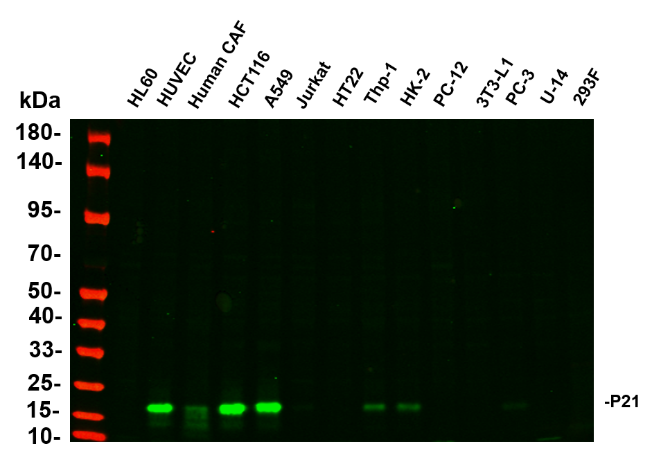
Various whole cell lysates were separated by 4-20% SDS-PAGE, and the primary antibody was used at 4℃, over night with a 1:5000 dilution . The Dylight 800-conjugated Goat anti-Rabbit antibody was used to detect the antibody. Lane1: HL60 - Human promyelocytic leukemia cell Lane2: HUVEC - Human umbilical vein endothelial cell Lane3: Human CAF - Human cancer-associated fibroblast Lane4: HCT116 - Human colorectal carcinoma Lane5: A549 - Human lung carcinoma Lane6: Jurkat - Human T lymphocyte leukemia Lane7: HT22 - Mouse hippocampal neuronal Lane8: Thp-1 - Human monocytic leukemia Lane9: HK-2 - Human proximal tubular epithelial Lane10: PC-12 - Rat adrenal pheochromocytoma Lane11: 3T3-L1 - Mouse embryonic fibroblast Lane12: PC-3 - Human prostate adenocarcinoma Lane13: U-14 - Mouse cervical carcinoma Lane14: 293F - HEK293 derivative, adapted for suspension culture.WB
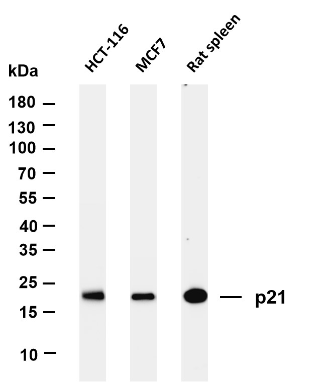
Various whole cell lysates were separated by 4-20% SDS-PAGE, and the membrane was blotted with anti-p21 antibody. The HRP-conjugated Goat anti-Rabbit IgG(H + L) antibody was used to detect the antibody. Lane 1: HCT-116 Lane 2: MCF7 Lane 3: Rat spleen.IHC
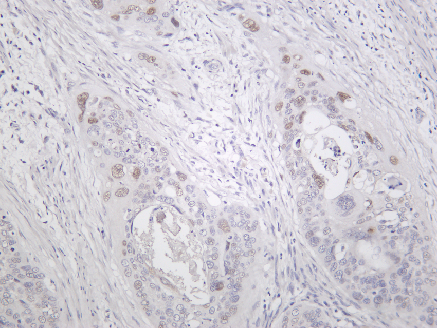
Human cervical carcinoma was stained with Anti-p21 rabbit antibody.ICC/IF
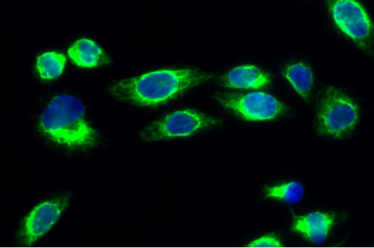
Immunofluorescence analysis of Hela cell. 1,p21 Antibody(green) was diluted at 1:200(4° overnight). 2, Goat Anti Rabbit Alexa Fluor 488 Catalog:RS3211 was diluted at 1:1000(room temperature, 50min). 3 DAPI(blue) 10min.ICC/IF
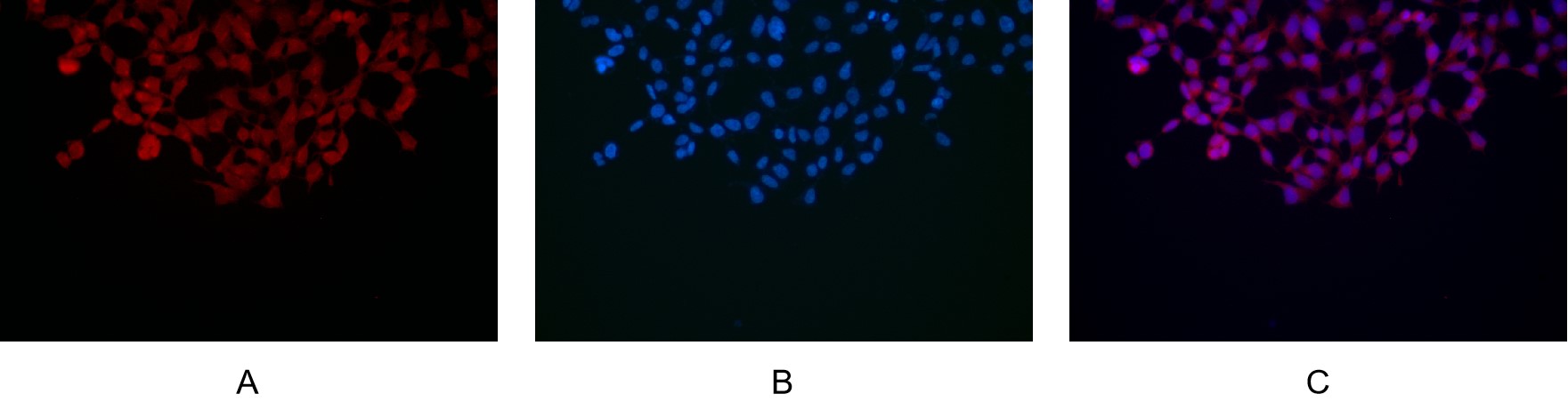
Immunofluorescence analysis of HEK293. Picture A: p21 antibody (red). Picture B: DAPI (blue). Picture C: Merge of A+B.| Product Name | p21 Rabbit mAb |
|---|---|
| Antibody Type | Primary Antibodies |
| Clonality | monoclonal |
|---|---|
| Isotype | IgG,K |
| Host Species | Rabbit |
| Tested Applications | ICC/IFIHCWB |
| WB: 1:1000-1:5000 IHC:1:100-1:200 ICC/IF:1:200-1:1000 |
|
| Species Reactivity | BovineHumanRat |
| Concentration | 1mg/ml |
| Purification | Protein A |
| Gene Symbol | CDKN1A |
|---|---|
| Gene Synonyms | P21 CIP1 SDI1 WAF1 CAP20 CDKN1 MDA-6 p21CIP1 |
| Gene Full Name | cyclin dependent kinase inhibitor 1A |
| Gene Summary | This gene encodes a potent cyclin-dependent kinase inhibitor. The encoded protein binds to and inhibits the activity of cyclin-cyclin-dependent kinase2 or -cyclin-dependent kinase4 complexes, and thus functions as a regulator of cell cycle progression at G1. The expression of this gene is tightly controlled by the tumor suppressor protein p53, through which this protein mediates the p53-dependent cell cycle G1 phase arrest in response to a variety of stress stimuli. This protein can interact with proliferating cell nuclear antigen, a DNA polymerase accessory factor, and plays a regulatory role in S phase DNA replication and DNA damage repair. This protein was reported to be specifically cleaved by CASP3-like caspases, which thus leads to a dramatic activation of cyclin-dependent kinase2, and may be instrumental in the execution of apoptosis following caspase activation. Mice that lack this gene have the ability to regenerate damaged or missing tissue. Multiple alternatively spliced variants have been found for this gene. [provided by RefSeq, Sep 2015] |
| Alternative Names | CDKN1A CAP20 CDKN1 CIP1 MDA6 PIC1 SDI1 WAF1 Cyclin-dependent kinase inhibitor 1 CDK-interacting protein 1 Melanoma differentiation-associated protein 6 MDA-6 p21 |
| Molecular Weight(MW) | 18kDa(Observed MW 18kDa) |
| Cellular Localization | Nucleus |
WB
Various whole cell lysates were separated by 4-20% SDS-PAGE, and the primary antibody was used at 4℃, over night with a 1:5000 dilution . The Dylight 800-conjugated Goat anti-Rabbit antibody was used to detect the antibody. Lane1: HL60 - Human promyelocytic leukemia cell Lane2: HUVEC - Human umbilical vein endothelial cell Lane3: Human CAF - Human cancer-associated fibroblast Lane4: HCT116 - Human colorectal carcinoma Lane5: A549 - Human lung carcinoma Lane6: Jurkat - Human T lymphocyte leukemia Lane7: HT22 - Mouse hippocampal neuronal Lane8: Thp-1 - Human monocytic leukemia Lane9: HK-2 - Human proximal tubular epithelial Lane10: PC-12 - Rat adrenal pheochromocytoma Lane11: 3T3-L1 - Mouse embryonic fibroblast Lane12: PC-3 - Human prostate adenocarcinoma Lane13: U-14 - Mouse cervical carcinoma Lane14: 293F - HEK293 derivative, adapted for suspension culture.
WB
Various whole cell lysates were separated by 4-20% SDS-PAGE, and the membrane was blotted with anti-p21 antibody. The HRP-conjugated Goat anti-Rabbit IgG(H + L) antibody was used to detect the antibody. Lane 1: HCT-116 Lane 2: MCF7 Lane 3: Rat spleen.
IHC
Human cervical carcinoma was stained with Anti-p21 rabbit antibody.
ICC/IF
Immunofluorescence analysis of Hela cell. 1,p21 Antibody(green) was diluted at 1:200(4° overnight). 2, Goat Anti Rabbit Alexa Fluor 488 Catalog:RS3211 was diluted at 1:1000(room temperature, 50min). 3 DAPI(blue) 10min.
ICC/IF
Immunofluorescence analysis of HEK293. Picture A: p21 antibody (red). Picture B: DAPI (blue). Picture C: Merge of A+B.| Application Notes | WB: 1:1000-1:5000 IHC:1:100-1:200 ICC/IF:1:200-1:1000 |
|---|
| Form | Liquid |
|---|---|
| Storage Instructions | -15°C to -25°C/1 year(Do not lower than -25°C) |
| Storage Buffer | PBS, 50% glycerol, 0.05% Proclin 300, 0.05%BSA |


