WB
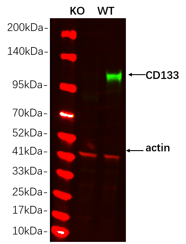
Western blot analysis of lysates from HAP1 WT and knockout cell , (Green) primary antibody was diluted at 1:5000, 4°over night, Dylight 800 secondary antibody was diluted at 1:10000, 37° 1hour. (Red) GAPDH Monoclonal Antibody(5B7) antibody was diluted at 1:5000 as loading control, 4° over night, Dylight 680 secondary antibody was diluted at 1:10000, 37° 1hour.WB
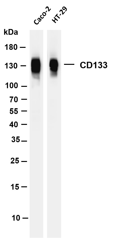
Various whole cell lysates were separated by 4-20% SDS-PAGE, and the membrane was blotted with anti-CD133 antibody. The HRP-conjugated Goat anti-Rabbit IgG(H + L) antibody was used to detect the antibody. Lane 1: Caco-2 Lane 2: HT-29.WB
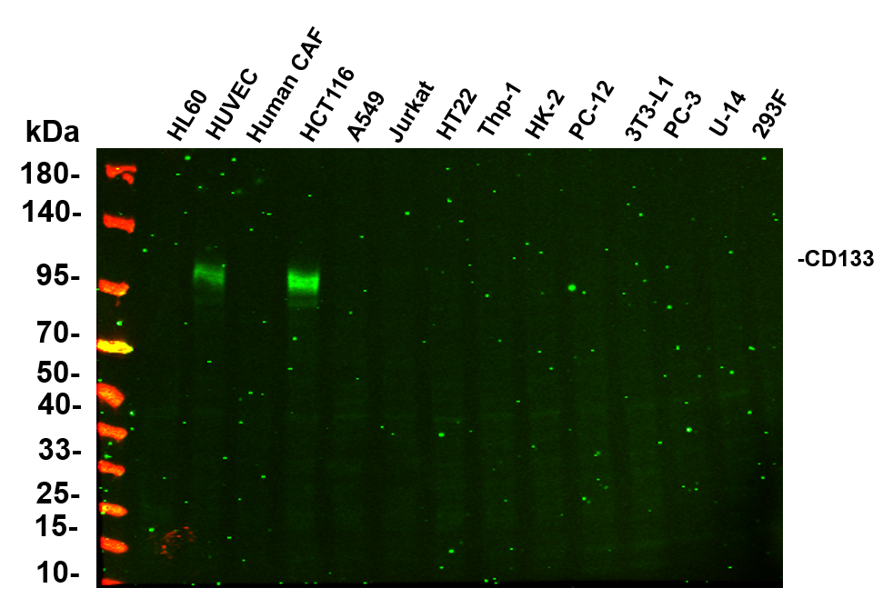
Various whole cell lysates were separated by 4-20% SDS-PAGE, and the primary antibody was used at 4℃, over night with a 1:2500 dilution . The Dylight 800-conjugated Goat anti-Rabbit antibody was used to detect the antibody. Lane1: HL60 - Human promyelocytic leukemia cell Lane2: HUVEC - Human umbilical vein endothelial cell Lane3: Human CAF - Human cancer-associated fibroblast Lane4: HCT116 - Human colorectal carcinoma Lane5: A549 - Human lung carcinoma Lane6: Jurkat - Human T lymphocyte leukemia Lane7: HT22 - Mouse hippocampal neuronal Lane8: Thp-1 - Human monocytic leukemia Lane9: HK-2 - Human proximal tubular epithelial Lane10: PC-12 - Rat adrenal pheochromocytoma Lane11: 3T3-L1 - Mouse embryonic fibroblast Lane12: PC-3 - Human prostate adenocarcinoma Lane13: U-14 - Mouse cervical carcinoma Lane14: 293F - HEK293 derivative, adapted for suspension culture.IHC
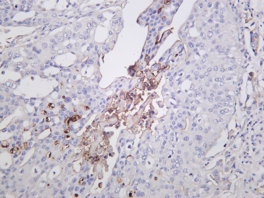
Human bladder carcinoma was stained with Anti-CD133 rabbit antibody.ICC/IF
Immunofluorescence analysis of A549. 1,primary Antibody(red) was diluted at 1:200(4℃ overnight). 2, Goat Anti Rabbit IgG (H&L) - Alexa Fluor 594 Secondary antibody was diluted at 1:1000(room temperature, 50min).3, DAPI(blue) 10min.| Product Name | CD133 Rabbit mAb |
|---|---|
| Antibody Type | Primary Antibodies |
| Clonality | monoclonal |
|---|---|
| Isotype | IgG,K |
| Host Species | Rabbit |
| Tested Applications | ICC/IFIHCWB |
| WB:1:1000-1:5000 IHC:1:20-100 ICC/IF:1:200-1:1000 |
|
| Species Reactivity | Human |
| Concentration | 1mg/ml |
| Purification | Protein A |
| Gene Symbol | PROM1 |
|---|---|
| Gene Synonyms | RP41 AC133 CD133 MCDR2 STGD4 CORD12 PROML1 MSTP061 |
| Gene Full Name | prominin 1 |
| Gene Summary | This gene encodes a pentaspan transmembrane glycoprotein. The protein localizes to membrane protrusions and is often expressed on adult stem cells, where it is thought to function in maintaining stem cell properties by suppressing differentiation. Mutations in this gene have been shown to result in retinitis pigmentosa and Stargardt disease. Expression of this gene is also associated with several types of cancer. This gene is expressed from at least five alternative promoters that are expressed in a tissue-dependent manner. Multiple transcript variants encoding different isoforms have been found for this gene. [provided by RefSeq, Mar 2009] |
| Alternative Names | PROM1 Prominin-1 Antigen AC133 Prominin-like protein 1 CD133 |
| Molecular Weight(MW) | 97kDa(Observed MW 133kDa) |
| Cellular Localization | Membranous |
WB
Western blot analysis of lysates from HAP1 WT and knockout cell , (Green) primary antibody was diluted at 1:5000, 4°over night, Dylight 800 secondary antibody was diluted at 1:10000, 37° 1hour. (Red) GAPDH Monoclonal Antibody(5B7) antibody was diluted at 1:5000 as loading control, 4° over night, Dylight 680 secondary antibody was diluted at 1:10000, 37° 1hour.
WB
Various whole cell lysates were separated by 4-20% SDS-PAGE, and the membrane was blotted with anti-CD133 antibody. The HRP-conjugated Goat anti-Rabbit IgG(H + L) antibody was used to detect the antibody. Lane 1: Caco-2 Lane 2: HT-29.
WB
Various whole cell lysates were separated by 4-20% SDS-PAGE, and the primary antibody was used at 4℃, over night with a 1:2500 dilution . The Dylight 800-conjugated Goat anti-Rabbit antibody was used to detect the antibody. Lane1: HL60 - Human promyelocytic leukemia cell Lane2: HUVEC - Human umbilical vein endothelial cell Lane3: Human CAF - Human cancer-associated fibroblast Lane4: HCT116 - Human colorectal carcinoma Lane5: A549 - Human lung carcinoma Lane6: Jurkat - Human T lymphocyte leukemia Lane7: HT22 - Mouse hippocampal neuronal Lane8: Thp-1 - Human monocytic leukemia Lane9: HK-2 - Human proximal tubular epithelial Lane10: PC-12 - Rat adrenal pheochromocytoma Lane11: 3T3-L1 - Mouse embryonic fibroblast Lane12: PC-3 - Human prostate adenocarcinoma Lane13: U-14 - Mouse cervical carcinoma Lane14: 293F - HEK293 derivative, adapted for suspension culture.
IHC
Human bladder carcinoma was stained with Anti-CD133 rabbit antibody.
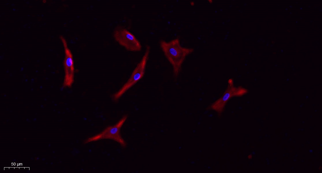
ICC/IF
Immunofluorescence analysis of A549. 1,primary Antibody(red) was diluted at 1:200(4℃ overnight). 2, Goat Anti Rabbit IgG (H&L) - Alexa Fluor 594 Secondary antibody was diluted at 1:1000(room temperature, 50min).3, DAPI(blue) 10min.| Application Notes | WB:1:1000-1:5000 IHC:1:20-100 ICC/IF:1:200-1:1000 |
|---|
| Form | Liquid |
|---|---|
| Storage Instructions | -15°C to -25°C/1 year(Do not lower than -25°C) |
| Storage Buffer | PBS, 50% glycerol, 0.05% Proclin 300, 0.05%BSA |


