WB
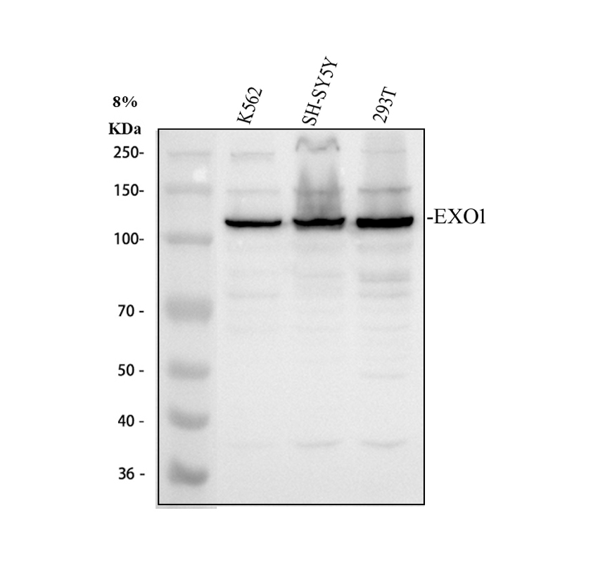
Western blot analysis of EXO1 using anti-EXO1 antibody. The sample well of each lane was loaded with 30 ug of sample under reducing conditions. Lane 1: human K562 whole cell lysates, Lane 2: human SH-SY5Y whole cell lysates, Lane 3: human 293T whole cell lysates. After electrophoresis, proteins were transferred to a membrane. Then the membrane was incubated with rabbit anti-EXO1 antigen affinity purified polyclonal antibody at a dilution of 1:1000 and probed with a goat anti-rabbit IgG-HRP secondary antibody. The signal is developed using ECL Plus Western Blotting Substrate.ICC/IF
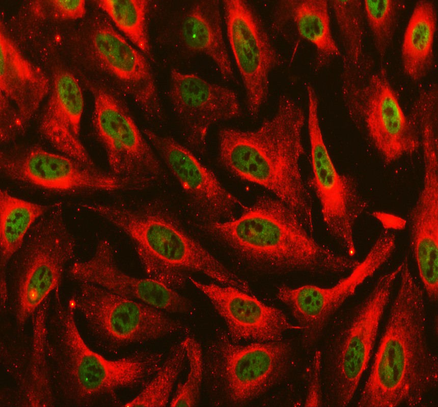
ICC/IF analysis of EXO1 using anti-EXO1 antibody and anti-Beta Tubulin antibody. EXO1 was detected in an immunocytochemical section of U2OS cells. The section was incubated with rabbit anti-EXO1 Antibody at a dilution of 1:100. Fluoro488-conjugated Anti-rabbit IgG Secondary Antibody (green) and Cy3-conjugated Anti-mouse IgG Secondary Antibody (red) were used as secondary antibody.IP
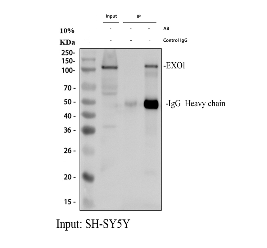
IP analysis of EXO1 using anti-EXO1 antibody in SH-SY5Y whole cell lysate. Western blot analysis of EXO1 using anti- EXO1 antibody. Lane 1: SH-SY5Y whole cell lysates(30ug), Lane 2: Rabbit control IgG instead of anti- EXO1 antibody in SH-SY5Y whole cell lysate, Lane 3: anti- EXO1 antibody (2μg) + SH-SY5Y whole cell lysate (500μg). After electrophoresis, proteins were transferred to a membrane. Then the membrane was incubated with rabbit anti- EXO1 antigen affinity purified polyclonal antibody at a dilution of 1:1000 and probed with a goat anti-rabbit IgG-HRP secondary antibody. The signal is developed using ECL Plus Western Blotting Substrate.| Product Name | Anti-EXO1 Antibody |
|---|---|
| Antibody Type | Primary Antibodies |
| Immunogen | E.coli-derived human Exonuclease 1/EXO1 recombinant protein (Position: Q25-Q846). |
| Clonality | Polyclonal |
|---|---|
| Isotype | IgG |
| Host Species | Rabbit |
| Tested Applications | ICC/IFIPWB |
| WB:1:500-2000 IP:1:250-300 ICC/IF:1:50-400 |
|
| Species Reactivity | Human |
| Concentration | 1mg/ml |
| Purification | Affinity purified |
| Gene Symbol | EXO1 |
|---|---|
| Gene Synonyms | HEX1 hExoI |
| Gene Full Name | exonuclease 1 |
| Gene Summary | This gene encodes a protein with 5' to 3' exonuclease activity as well as an RNase H activity. It is similar to the Saccharomyces cerevisiae protein Exo1 which interacts with Msh2 and which is involved in mismatch repair and recombination. Alternative splicing of this gene results in three transcript variants encoding two different isoforms. [provided by RefSeq, Jul 2008] |
| Alternative Names | EXO1 EXOI exonuclease 1 Exonuclease I HEX1 hExo1 hExoI |
| Molecular Weight(MW) | 94kDa(Observed MW 120kDa) |
| Cellular Localization | Nucleus |
WB
Western blot analysis of EXO1 using anti-EXO1 antibody. The sample well of each lane was loaded with 30 ug of sample under reducing conditions. Lane 1: human K562 whole cell lysates, Lane 2: human SH-SY5Y whole cell lysates, Lane 3: human 293T whole cell lysates. After electrophoresis, proteins were transferred to a membrane. Then the membrane was incubated with rabbit anti-EXO1 antigen affinity purified polyclonal antibody at a dilution of 1:1000 and probed with a goat anti-rabbit IgG-HRP secondary antibody. The signal is developed using ECL Plus Western Blotting Substrate.
ICC/IF
ICC/IF analysis of EXO1 using anti-EXO1 antibody and anti-Beta Tubulin antibody. EXO1 was detected in an immunocytochemical section of U2OS cells. The section was incubated with rabbit anti-EXO1 Antibody at a dilution of 1:100. Fluoro488-conjugated Anti-rabbit IgG Secondary Antibody (green) and Cy3-conjugated Anti-mouse IgG Secondary Antibody (red) were used as secondary antibody.
IP
IP analysis of EXO1 using anti-EXO1 antibody in SH-SY5Y whole cell lysate. Western blot analysis of EXO1 using anti- EXO1 antibody. Lane 1: SH-SY5Y whole cell lysates(30ug), Lane 2: Rabbit control IgG instead of anti- EXO1 antibody in SH-SY5Y whole cell lysate, Lane 3: anti- EXO1 antibody (2μg) + SH-SY5Y whole cell lysate (500μg). After electrophoresis, proteins were transferred to a membrane. Then the membrane was incubated with rabbit anti- EXO1 antigen affinity purified polyclonal antibody at a dilution of 1:1000 and probed with a goat anti-rabbit IgG-HRP secondary antibody. The signal is developed using ECL Plus Western Blotting Substrate.| Application Notes | WB:1:500-2000 IP:1:250-300 ICC/IF:1:50-400 |
|---|
| Form | Liquid |
|---|---|
| Storage Instructions | 12 months from date of receipt,-20℃ as supplied. |
| Storage Buffer | 500 ug/ml antibody with PBS, 0.02% NaN3, 1 mg/ml BSA and 50% glycerol. |


