
WB
Western Blot, Positive WB detected in: K562 whole cell lysate(30µg), A549 whole cell lysate(30µg), Rat Liver tissue lysate(30µg),Rat Lung tissue lysate(30µg), All lanes: HBB antibody at 1:1000, Secondary, Goat polyclonal to rabbit IgG at 1/40000 dilution, Exposure time: 120s.IHC
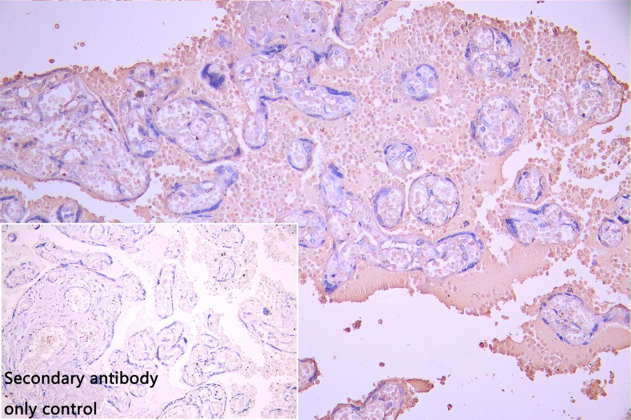
IHC image of HBB Antibody diluted at 1:100 and staining in paraffinembedded human placenta tissue performed ona Leica BondTM system. After dewaxing and hydration, antigen retrieval was mediated by high pressure in a citrate buffer (pH 6.0). Section was blocked with 10% normal goat serum 30min at RT. Then primary antibody (1% BSA) was incubated at 4°C overnight. The primary is detected by a Goat antirabbit polymer IgG labeled by HRP and visualized using 0.05% DAB.Secondary antibody only control: uses 1% BSAinstead of primary antibody.| Product Name | HBB Antibody |
|---|---|
| Antibody Type | Primary Antibodies |
| Immunogen | Recombinant Human Hemoglobin subunit beta protein (1-147AA) |
| Clonality | Polyclonal |
|---|---|
| Isotype | IgG |
| Host Species | Rabbit |
| Tested Applications | IHCWB |
| WB:1:500-1:2000 IHC:1:50-1:200 |
|
| Species Reactivity | HumanRat |
| Concentration | 1mg/ml |
| Purification | Protein G |
| Gene Symbol | HBB |
|---|---|
| Gene Synonyms | ECYT6 CD113t-C beta-globin |
| Gene Full Name | hemoglobin subunit beta |
| Gene Summary | The alpha (HBA) and beta (HBB) loci determine the structure of the 2 types of polypeptide chains in adult hemoglobin, Hb A. The normal adult hemoglobin tetramer consists of two alpha chains and two beta chains. Mutant beta globin causes sickle cell anemia. Absence of beta chain causes beta-zero-thalassemia. Reduced amounts of detectable beta globin causes beta-plus-thalassemia. The order of the genes in the beta-globin cluster is 5'-epsilon -- gamma-G -- gamma-A -- delta -- beta--3'. [provided by RefSeq, Jul 2008] |
| Molecular Weight(MW) | 16kDa(Observed MW 16kDa) |
WB
Western Blot, Positive WB detected in: K562 whole cell lysate(30µg), A549 whole cell lysate(30µg), Rat Liver tissue lysate(30µg),Rat Lung tissue lysate(30µg), All lanes: HBB antibody at 1:1000, Secondary, Goat polyclonal to rabbit IgG at 1/40000 dilution, Exposure time: 120s.
IHC
IHC image of HBB Antibody diluted at 1:100 and staining in paraffinembedded human placenta tissue performed ona Leica BondTM system. After dewaxing and hydration, antigen retrieval was mediated by high pressure in a citrate buffer (pH 6.0). Section was blocked with 10% normal goat serum 30min at RT. Then primary antibody (1% BSA) was incubated at 4°C overnight. The primary is detected by a Goat antirabbit polymer IgG labeled by HRP and visualized using 0.05% DAB.Secondary antibody only control: uses 1% BSAinstead of primary antibody.| Application Notes | WB:1:500-1:2000 IHC:1:50-1:200 |
|---|
| Form | Liquid |
|---|---|
| Storage Instructions | Upon receipt, store at -20°C or -80°C. Avoid repeated freeze. |
| Storage Buffer | Preservative: 0.03% Proclin 300 Constituents: 50% Glycerol, 0.01M PBS, pH 7.4 |


