WB
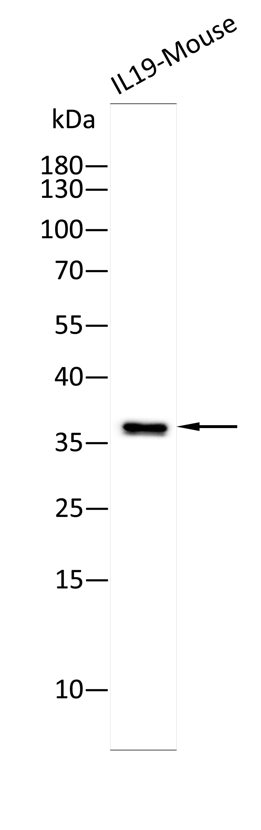
Western blot analysis using Anti-IL19-Mouse antibody against Recombinant IL19-Mouse protein. 15% SDS-PAGE gel.Sample loading: 10ng /lane. Transfer the proteins onto a PVDF membrane (OM790002), and block it with TBST (OM750016) plus skimmed milk powder(OM750084) for one hour. Dilute the primary antibody with the antibody diluent (OM750012) at a ratio of 1:5000, and incubate it overnight at 4°C. Wash the membrane three times with TBST (OM750016), 5 minutes each time. At room temperature, dilute the secondary antibody, Goat Anti-Rabbit IgG(H&L)-HRP (OM643487), at a ratio of 1:10000 and incubate for one hour. Wash the membrane three times with TBST (OM750016) again, 5 minutes each time. Use ECL (OM625701) for luminescence.staining time: 30S.ELISA
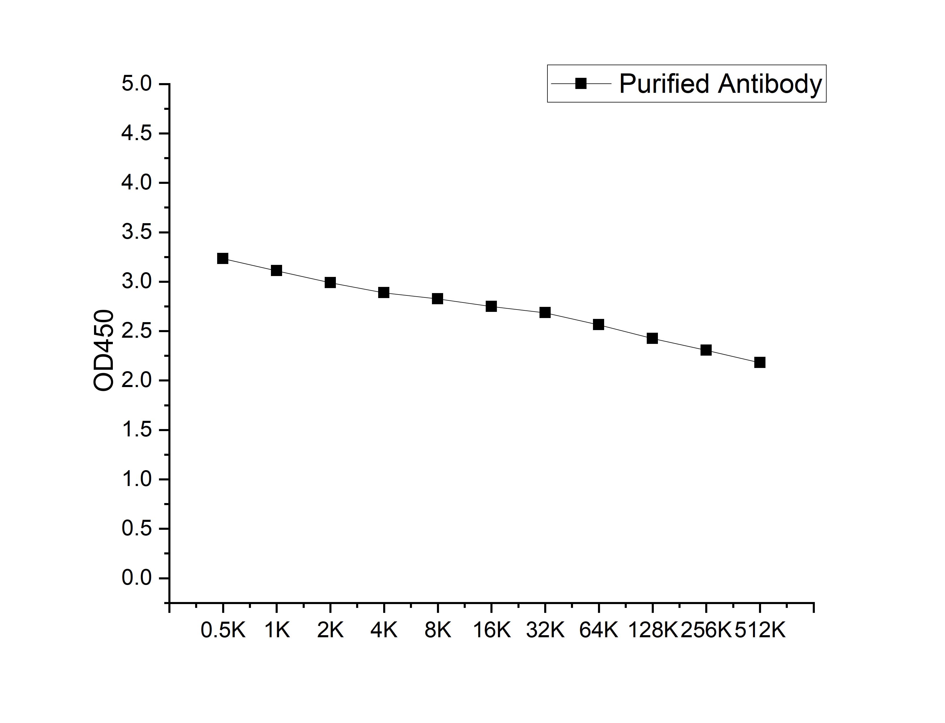
Microtiter plate wells were coated with 100 ng/well coating antigen and incubated with serial dilutions of the antibody. Following HRP-conjugated secondary antibody and substrate development, OD450 was measured. The absorbance decreased progressively with increasing antibody dilution, yielding a sigmoidal titration curve that demonstrates the high antigen-binding activity of the polyclonal antibody.ICC/IF
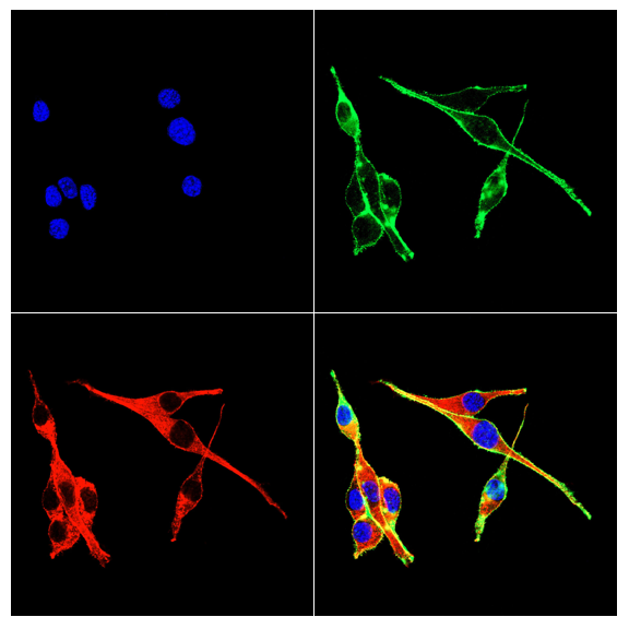
Immunofluorescence analysis of NIH3T3 cells using IL19 antibody (green). Blue: DAPI fluorescent DNA dye. Red: Actin filaments have been labeled with Omnimabs® 594-Phalloidin.Cells are fixed in 4% paraformaldehyde at room temperature for 15 minutes. Then, they are permeabilized with a PBS (OM750003) solution containing 0.1% Triton X-100(OM750021) at room temperature for 15 minutes. Subsequently, the cells are blocked with 10% non - immune goat serum(OM760028) at room temperature for 1 hour.The cells are incubated overnight at 4°C with the primary antibody diluted 1:100 in PBS. The secondary antibody, Omnimabs® 488 Goat Ant-Rabbit IgG(H&L) (Green,OM643486), is diluted at a ratio of 1:400 and incubated with the cells for 1 hour.Nuclear DNA is labeled with DAPI (Blue,OM643160). F-actin is stained with Omnimabs® 594-Phalloidin (Red,OM750007) diluted 1:100 for 30 minutes.| Product Name | Anti-IL19 antibody |
|---|---|
| Antibody Type | Primary Antibodies |
| Immunogen | Recombinant Mouse IL19 protein |
| Clonality | polyclonal |
|---|---|
| Isotype | IgG |
| Host Species | Rabbit |
| Tested Applications | ELISAICC/IFWB |
| WB:1:5000 ICC/IF:1:100-1:500 |
|
| Species Reactivity | Mouse |
| Concentration | 1mg/ml |
| Purification | Affinity purified |
| Gene Symbol | IL19 |
|---|---|
| Gene Synonyms | IL-10C MDA1 NG.1 ZMDA1 |
| Gene Full Name | interleukin 19 |
| Gene Summary | The protein encoded by this gene is a cytokine that belongs to the IL10 cytokine subfamily. This cytokine is found to be preferentially expressed in monocytes. It can bind the IL20 receptor complex and lead to the activation of the signal transducer and activator of transcription 3 (STAT3). A similar cytokine in mouse is reported to up-regulate the expression of IL6 and TNF-alpha and induce apoptosis, which suggests a role of this cytokine in inflammatory responses. Alternatively spliced transcript variants encoding the distinct isoforms have been described. [provided by RefSeq, Jul 2008] |
| Alternative Names | cytokine il 19 IL-19 ILN Interleukin Interleukin19 MDA-1 melanoma differentiation associated protein-like protein ZMDA1 |
| Cellular Localization | Secreted. |
| SwissProt ID | Q8CJ70 |
|---|
WB
Western blot analysis using Anti-IL19-Mouse antibody against Recombinant IL19-Mouse protein. 15% SDS-PAGE gel.Sample loading: 10ng /lane. Transfer the proteins onto a PVDF membrane (OM790002), and block it with TBST (OM750016) plus skimmed milk powder(OM750084) for one hour. Dilute the primary antibody with the antibody diluent (OM750012) at a ratio of 1:5000, and incubate it overnight at 4°C. Wash the membrane three times with TBST (OM750016), 5 minutes each time. At room temperature, dilute the secondary antibody, Goat Anti-Rabbit IgG(H&L)-HRP (OM643487), at a ratio of 1:10000 and incubate for one hour. Wash the membrane three times with TBST (OM750016) again, 5 minutes each time. Use ECL (OM625701) for luminescence.staining time: 30S.
ELISA
Microtiter plate wells were coated with 100 ng/well coating antigen and incubated with serial dilutions of the antibody. Following HRP-conjugated secondary antibody and substrate development, OD450 was measured. The absorbance decreased progressively with increasing antibody dilution, yielding a sigmoidal titration curve that demonstrates the high antigen-binding activity of the polyclonal antibody.
ICC/IF
Immunofluorescence analysis of NIH3T3 cells using IL19 antibody (green). Blue: DAPI fluorescent DNA dye. Red: Actin filaments have been labeled with Omnimabs® 594-Phalloidin.Cells are fixed in 4% paraformaldehyde at room temperature for 15 minutes. Then, they are permeabilized with a PBS (OM750003) solution containing 0.1% Triton X-100(OM750021) at room temperature for 15 minutes. Subsequently, the cells are blocked with 10% non - immune goat serum(OM760028) at room temperature for 1 hour.The cells are incubated overnight at 4°C with the primary antibody diluted 1:100 in PBS. The secondary antibody, Omnimabs® 488 Goat Ant-Rabbit IgG(H&L) (Green,OM643486), is diluted at a ratio of 1:400 and incubated with the cells for 1 hour.Nuclear DNA is labeled with DAPI (Blue,OM643160). F-actin is stained with Omnimabs® 594-Phalloidin (Red,OM750007) diluted 1:100 for 30 minutes.| Application Notes | WB:1:5000 ICC/IF:1:100-1:500 |
|---|
| Form | Liquid |
|---|---|
| Storage Instructions | Store at 4°C short term. Aliquot and store at -20°C long term. Avoid freeze/thaw cycles. |
| Storage Buffer | Purified antibody in PBS with 0.05% sodium azide,40% Glycerol. |


