WB
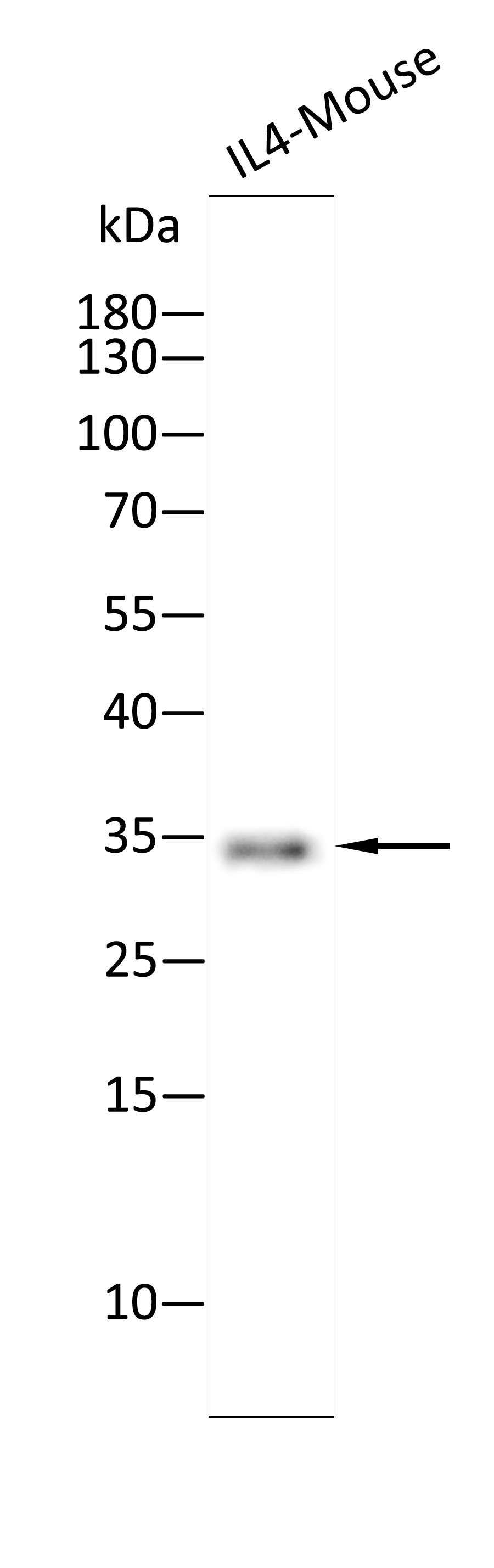
Western blot analysis using Anti-IL4-Mouse antibody against Recombinant IL4-Mouse protein. 15% SDS-PAGE gel.Sample loading: 10ng /lane. Transfer the proteins onto a PVDF membrane (OM790002), and block it with TBST (OM750016) plus skimmed milk powder(OM750084) for one hour. Dilute the primary antibody with the antibody diluent (OM750012) at a ratio of 1:5000, and incubate it overnight at 4°C. Wash the membrane three times with TBST (OM750016), 5 minutes each time. At room temperature, dilute the secondary antibody, Goat Anti-Rabbit IgG(H&L)-HRP (OM643487), at a ratio of 1:10000 and incubate for one hour. Wash the membrane three times with TBST (OM750016) again, 5 minutes each time. Use ECL (OM625701) for luminescence.staining time: 30S.WB
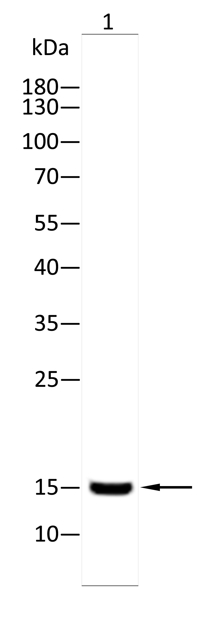
Western blot analysis using IL4 antibody against Hepg2(1) cell . 12% SDS-PAGE gel.Sample loading: 20μg /lane. Transfer the proteins onto a PVDF membrane (OM790003), and block it with TBST (OM750016) plus skimmed milk powder(OM750084) for one hour. Dilute the primary antibody with the antibody diluent (OM750012) at a ratio of 1:1000, and incubate it overnight at 4°C. Wash the membrane three times with TBST (OM750016), 5 minutes each time. At room temperature, dilute the secondary antibody, Goat Anti-Rabbit IgG(H&L)-HRP (OM643487), at a ratio of 1:20000 and incubate for one hour. Wash the membrane three times with TBST (OM750016) again, 5 minutes each time. Use ECL (OM625701) for luminescence.staining time: 60S.ELISA
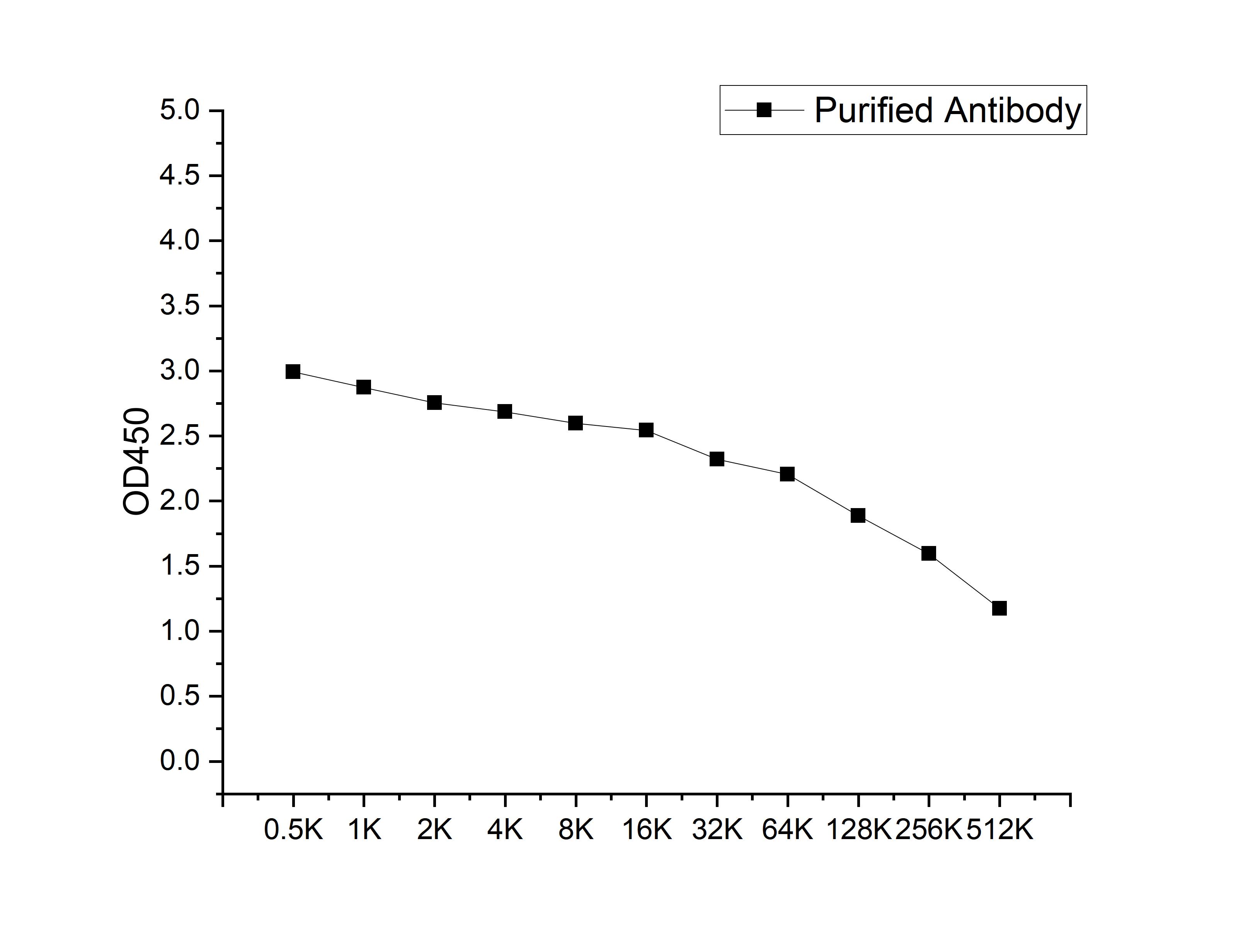
Microtiter plate wells were coated with 100 ng/well coating antigen and incubated with serial dilutions of the antibody. Following HRP-conjugated secondary antibody and substrate development, OD450 was measured. The absorbance decreased progressively with increasing antibody dilution, yielding a sigmoidal titration curve that demonstrates the high antigen-binding activity of the polyclonal antibody.| Product Name | Anti-IL4 antibody |
|---|---|
| Antibody Type | Primary Antibodies |
| Immunogen | Recombinant Mouse IL4 protein |
| Clonality | polyclonal |
|---|---|
| Isotype | IgG |
| Host Species | Rabbit |
| Tested Applications | ELISAWB |
| WB:1:1000-1:2000 |
|
| Species Reactivity | HumanMouse |
| Concentration | 1mg/ml |
| Purification | Affinity purified |
| Gene Symbol | IL4 |
|---|---|
| Gene Synonyms | BCGF-1 BCGF1 BSF-1 BSF1 IL-4 Il4e12 |
| Gene Full Name | interleukin 4 |
| Gene Summary | The protein encoded by this gene is a pleiotropic cytokine produced by activated T cells. This cytokine is a ligand for interleukin 4 receptor. The interleukin 4 receptor also binds to IL13, which may contribute to many overlapping functions of this cytokine and IL13. STAT6, a signal transducer and activator of transcription, has been shown to play a central role in mediating the immune regulatory signal of this cytokine. This gene, IL3, IL5, IL13, and CSF2 form a cytokine gene cluster on chromosome 5q, with this gene particularly close to IL13. This gene, IL13 and IL5 are found to be regulated coordinately by several long-range regulatory elements in an over 120 kilobase range on the chromosome. IL4 is considered an important cytokine for tissue repair, counterbalancing the effects of proinflammatory type 1 cytokines, however, it also promotes allergic airway inflammation. Moreover, IL-4, a type 2 cytokine, mediates and regulates a variety of human host responses such as allergic, anti-parasitic, wound healing, and acute inflammation. This cytokine has been reported to promote resolution of neutrophil-mediated acute lung injury. In an allergic response, IL-4 has an essential role in the production of allergen-specific immunoglobin (Ig) E. This pro-inflammatory cytokine has been observed to be increased in COVID-19 (Coronavirus disease 2019) patients, but is not necessarily associated with severe COVID-19 pathology. Two alternatively spliced transcript variants of this gene encoding distinct isoforms have been reported. [provided by RefSeq, Aug 2020] |
| Alternative Names | B cell growth factor 1 B-cell growth factor 1 B-cell IGG differentiation factor B-cell stimulatory factor 1 B_cell stimulatory factor 1 binetrakin BSF-1 H-IL-4 IGG1 induction factor IL-4 IL-4 cytokine IL-4v2 IL4 ILN Interleukin interleukin 4 interleukin mRNA Interleukin4 lymphocyte stimulatory factor 1 M-IL-4 MGC79402 pitrakinra |
| Molecular Weight(MW) | 16kDa(observed:34、15kDa) |
| Cellular Localization | Secreted. |
| SwissProt ID | P07750 |
|---|
WB
Western blot analysis using Anti-IL4-Mouse antibody against Recombinant IL4-Mouse protein. 15% SDS-PAGE gel.Sample loading: 10ng /lane. Transfer the proteins onto a PVDF membrane (OM790002), and block it with TBST (OM750016) plus skimmed milk powder(OM750084) for one hour. Dilute the primary antibody with the antibody diluent (OM750012) at a ratio of 1:5000, and incubate it overnight at 4°C. Wash the membrane three times with TBST (OM750016), 5 minutes each time. At room temperature, dilute the secondary antibody, Goat Anti-Rabbit IgG(H&L)-HRP (OM643487), at a ratio of 1:10000 and incubate for one hour. Wash the membrane three times with TBST (OM750016) again, 5 minutes each time. Use ECL (OM625701) for luminescence.staining time: 30S.
WB
Western blot analysis using IL4 antibody against Hepg2(1) cell . 12% SDS-PAGE gel.Sample loading: 20μg /lane. Transfer the proteins onto a PVDF membrane (OM790003), and block it with TBST (OM750016) plus skimmed milk powder(OM750084) for one hour. Dilute the primary antibody with the antibody diluent (OM750012) at a ratio of 1:1000, and incubate it overnight at 4°C. Wash the membrane three times with TBST (OM750016), 5 minutes each time. At room temperature, dilute the secondary antibody, Goat Anti-Rabbit IgG(H&L)-HRP (OM643487), at a ratio of 1:20000 and incubate for one hour. Wash the membrane three times with TBST (OM750016) again, 5 minutes each time. Use ECL (OM625701) for luminescence.staining time: 60S.
ELISA
Microtiter plate wells were coated with 100 ng/well coating antigen and incubated with serial dilutions of the antibody. Following HRP-conjugated secondary antibody and substrate development, OD450 was measured. The absorbance decreased progressively with increasing antibody dilution, yielding a sigmoidal titration curve that demonstrates the high antigen-binding activity of the polyclonal antibody.| Application Notes | WB:1:1000-1:2000 |
|---|
| Form | Liquid |
|---|---|
| Storage Instructions | Store at 4°C short term. Aliquot and store at -20°C long term. Avoid freeze/thaw cycles. |
| Storage Buffer | Purified antibody in PBS with 0.05% sodium azide,40% Glycerol. |


