WB
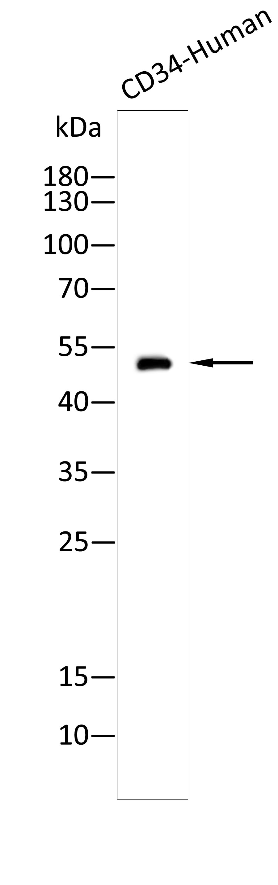
Western blot analysis using Anti-CD34-Human antibody against Recombinant CD34-Human protein. 12% SDS-PAGE gel.Sample loading: 10ng /lane. Transfer the proteins onto a PVDF membrane (OM790002), and block it with TBST (OM750016) plus skimmed milk powder(OM750084) for one hour. Dilute the primary antibody with the antibody diluent (OM750012) at a ratio of 1:5000, and incubate it overnight at 4°C. Wash the membrane three times with TBST (OM750016), 5 minutes each time. At room temperature, dilute the secondary antibody, Goat Anti-Rabbit IgG(H&L)-HRP (OM643487), at a ratio of 1:10000 and incubate for one hour. Wash the membrane three times with TBST (OM750016) again, 5 minutes each time. Use ECL (OM625701) for luminescence.staining time: 30S.WB
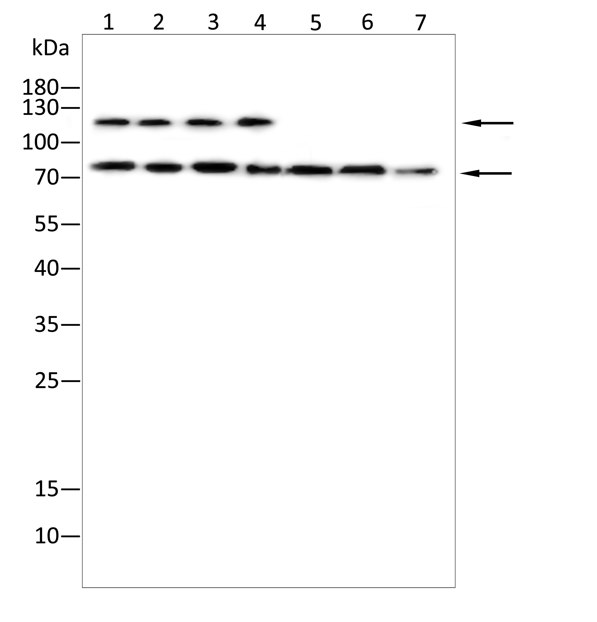
Western blot analysis using CD34 antibody against Hela(1),Cos-7(2),K562(3),Hepg2(4),A549(5),Nih3t3(6),Mouse liver tissue(7) . 8% SDS-PAGE gel.Sample loading: 20μg /lane. Transfer the proteins onto a PVDF membrane (OM790003), and block it with TBST (OM750016) plus skimmed milk powder(OM750084) for one hour. Dilute the primary antibody with the antibody diluent (OM750012) at a ratio of 1:1000, and incubate it overnight at 4°C. Wash the membrane three times with TBST (OM750016), 5 minutes each time. At room temperature, dilute the secondary antibody, Goat Anti-Rabbit IgG(H&L)-HRP (OM643487), at a ratio of 1:20000 and incubate for one hour. Wash the membrane three times with TBST (OM750016) again, 5 minutes each time. Use ECL (OM625701) for luminescence.staining time: 60S.ELISA
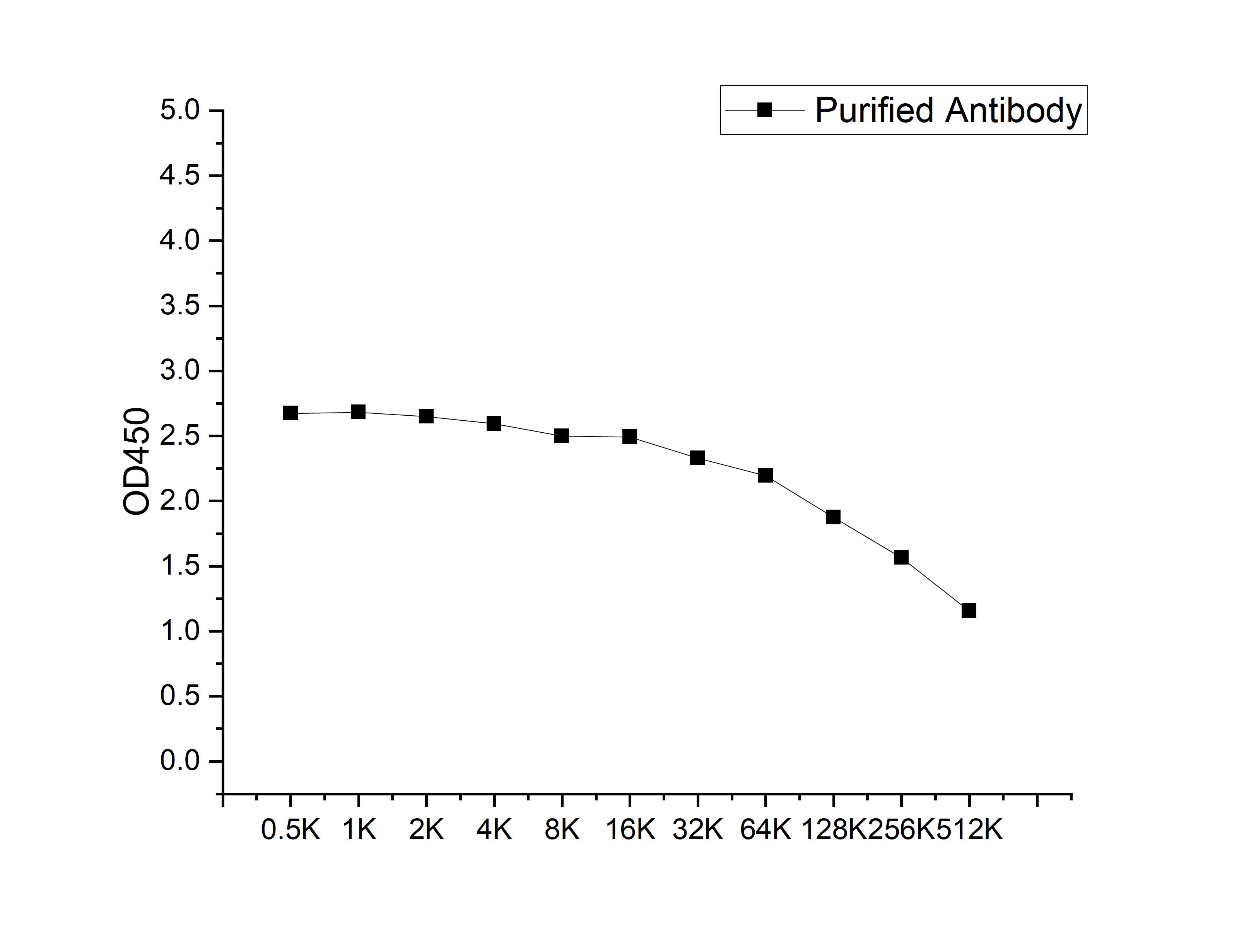
Microtiter plate wells were coated with 100 ng/well coating antigen and incubated with serial dilutions of the antibody. Following HRP-conjugated secondary antibody and substrate development, OD450 was measured. The absorbance decreased progressively with increasing antibody dilution, yielding a sigmoidal titration curve that demonstrates the high antigen-binding activity of the polyclonal antibody.| Product Name | Anti-CD34 antibody |
|---|---|
| Antibody Type | Primary Antibodies |
| Immunogen | Recombinant Human CD34 protein |
| Clonality | polyclonal |
|---|---|
| Isotype | IgG |
| Host Species | Rabbit |
| Tested Applications | ELISAWB |
| WB:1:1000-1:2000 |
|
| Species Reactivity | HumanMouse |
| Concentration | 1mg/ml |
| Purification | Affinity purified |
| Gene Symbol | CD34 |
|---|---|
| Gene Full Name | CD34 molecule |
| Gene Summary | The protein encoded by this gene may play a role in the attachment of stem cells to the bone marrow extracellular matrix or to stromal cells. This single-pass membrane protein is highly glycosylated and phosphorylated by protein kinase C. Two transcript variants encoding different isoforms have been found for this gene. [provided by RefSeq, Aug 2011] |
| Alternative Names | CD34 antigen CD34 sialomucin cell surface antigen cluster designation 34 glycoprotein expressed in lymphohematopoietic progenitor cells hematopoietic progenitor cell marker CD34 Mucosialin RP11-328D5.2 sialomucin protein stem cell antigen type 1 transmembrane protein |
| Molecular Weight(MW) | 41kDa(observed:53、80/115kDa) |
| Cellular Localization | Membrane. |
| SwissProt ID | P28906 |
|---|
WB
Western blot analysis using Anti-CD34-Human antibody against Recombinant CD34-Human protein. 12% SDS-PAGE gel.Sample loading: 10ng /lane. Transfer the proteins onto a PVDF membrane (OM790002), and block it with TBST (OM750016) plus skimmed milk powder(OM750084) for one hour. Dilute the primary antibody with the antibody diluent (OM750012) at a ratio of 1:5000, and incubate it overnight at 4°C. Wash the membrane three times with TBST (OM750016), 5 minutes each time. At room temperature, dilute the secondary antibody, Goat Anti-Rabbit IgG(H&L)-HRP (OM643487), at a ratio of 1:10000 and incubate for one hour. Wash the membrane three times with TBST (OM750016) again, 5 minutes each time. Use ECL (OM625701) for luminescence.staining time: 30S.
WB
Western blot analysis using CD34 antibody against Hela(1),Cos-7(2),K562(3),Hepg2(4),A549(5),Nih3t3(6),Mouse liver tissue(7) . 8% SDS-PAGE gel.Sample loading: 20μg /lane. Transfer the proteins onto a PVDF membrane (OM790003), and block it with TBST (OM750016) plus skimmed milk powder(OM750084) for one hour. Dilute the primary antibody with the antibody diluent (OM750012) at a ratio of 1:1000, and incubate it overnight at 4°C. Wash the membrane three times with TBST (OM750016), 5 minutes each time. At room temperature, dilute the secondary antibody, Goat Anti-Rabbit IgG(H&L)-HRP (OM643487), at a ratio of 1:20000 and incubate for one hour. Wash the membrane three times with TBST (OM750016) again, 5 minutes each time. Use ECL (OM625701) for luminescence.staining time: 60S.
ELISA
Microtiter plate wells were coated with 100 ng/well coating antigen and incubated with serial dilutions of the antibody. Following HRP-conjugated secondary antibody and substrate development, OD450 was measured. The absorbance decreased progressively with increasing antibody dilution, yielding a sigmoidal titration curve that demonstrates the high antigen-binding activity of the polyclonal antibody.| Application Notes | WB:1:1000-1:2000 |
|---|
| Form | Liquid |
|---|---|
| Storage Instructions | Store at 4°C short term. Aliquot and store at -20°C long term. Avoid freeze/thaw cycles. |
| Storage Buffer | Purified antibody in PBS with 0.05% sodium azide,40% Glycerol. |


