WB
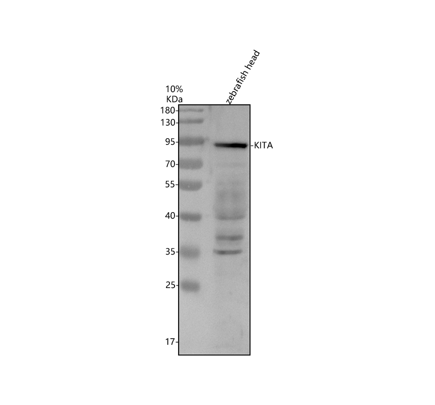
Western blot analysis of KITA using anti-KITA antibody . The sample well of each lane was loaded with 30 ug of sample under reducing conditions.Lane 1: zebrafish head tissue lysates.After electrophoresis, proteins were transferred to a membrane. Then the membrane was incubated with rabbit anti-KITA antigen affinity purified polyclonal antibody at a dilution of 1:1000 and probed with a goat anti-rabbit IgG-HRP secondary antibody . The signal is developed using ECL Plus Western Blotting Substrate . A specific band was detected for KITA at approximately 95 kDa. The expected band size for KITA is at 109 kDa.IHC
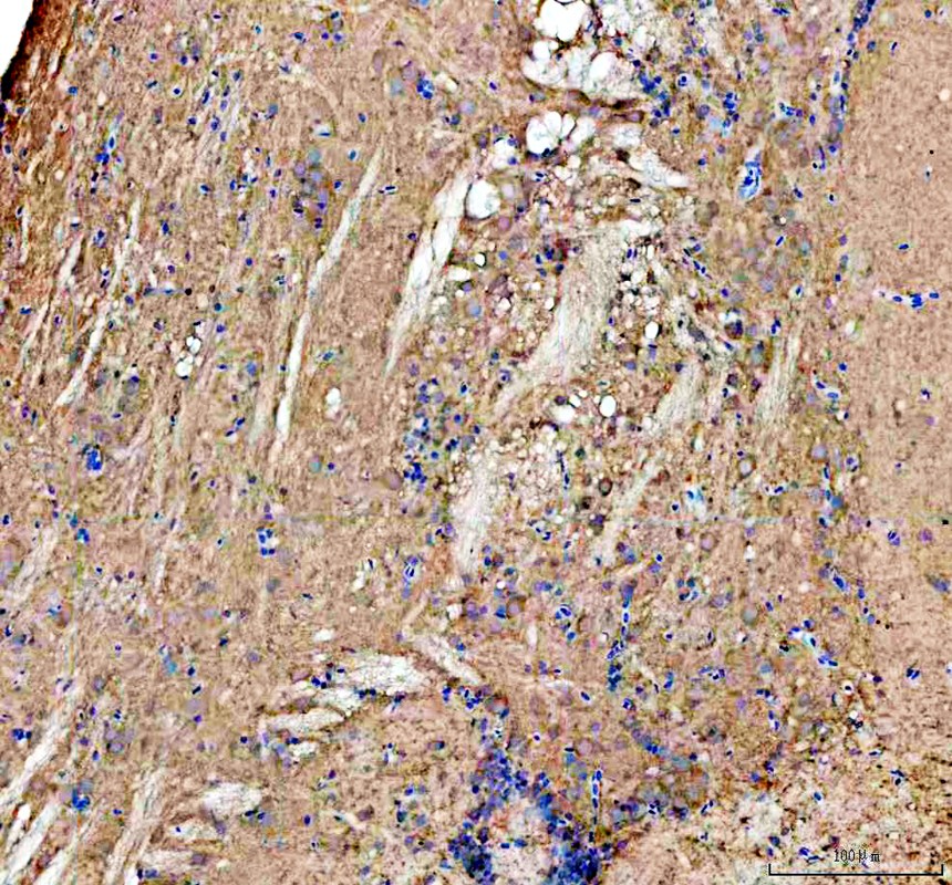
IHC analysis of KITA using anti-KITA antibody .KITA was detected in a paraffin-embedded section of zebrafish brain tissue. The tissue section was incubated with rabbit anti-KITA Antibody at a dilution of 1:200 and developed using HRP Conjugated Rabbit IgG Super Vision Assay Kit with DAB as the chromogen.IHC
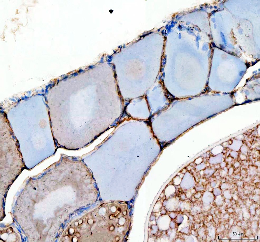
IHC analysis of KITA using anti-KITA antibody .KITA was detected in a paraffin-embedded section of zebrafish ovary tissue. The tissue section was incubated with rabbit anti-KITA Antibody at a dilution of 1:200 and developed using HRP Conjugated Rabbit IgG Super Vision Assay Kit with DAB as the chromogen.| Product Name | Anti-Zebrafish KITA Antibody |
|---|---|
| Antibody Type | Primary Antibodies |
| Immunogen | E.coli-derived Zebrafish KITA recombinant protein (Position: R22-N946) |
| Clonality | Polyclonal |
|---|---|
| Isotype | IgG |
| Host Species | Rabbit |
| Tested Applications | WBIHC |
| WB:1:500-1:2000 IHC:1:50-1:400 |
|
| Species Reactivity | Zebrafish |
| Concentration | 500ug/ml |
| Purification | Affinity purified |
| Gene Full Name | KIT proto-oncogene, receptor tyrosine kinase a |
|---|---|
| Target | KITA |
| Alternative Names | kita kit sparse |
| Molecular Weight(MW) | 109 kDa(Observed band size: 95 kDa) |
| SwissProt ID | Q8JFR5 |
|---|
WB
Western blot analysis of KITA using anti-KITA antibody . The sample well of each lane was loaded with 30 ug of sample under reducing conditions.Lane 1: zebrafish head tissue lysates.After electrophoresis, proteins were transferred to a membrane. Then the membrane was incubated with rabbit anti-KITA antigen affinity purified polyclonal antibody at a dilution of 1:1000 and probed with a goat anti-rabbit IgG-HRP secondary antibody . The signal is developed using ECL Plus Western Blotting Substrate . A specific band was detected for KITA at approximately 95 kDa. The expected band size for KITA is at 109 kDa.
IHC
IHC analysis of KITA using anti-KITA antibody .KITA was detected in a paraffin-embedded section of zebrafish brain tissue. The tissue section was incubated with rabbit anti-KITA Antibody at a dilution of 1:200 and developed using HRP Conjugated Rabbit IgG Super Vision Assay Kit with DAB as the chromogen.
IHC
IHC analysis of KITA using anti-KITA antibody .KITA was detected in a paraffin-embedded section of zebrafish ovary tissue. The tissue section was incubated with rabbit anti-KITA Antibody at a dilution of 1:200 and developed using HRP Conjugated Rabbit IgG Super Vision Assay Kit with DAB as the chromogen.| Application Notes | WB:1:500-1:2000 IHC:1:50-1:400 |
|---|
| Form | Liquid |
|---|---|
| Storage Instructions | 12 months from date of receipt,-20℃ as supplied. |
| Storage Buffer | PBS, 0.02% NaN3, 1 mg/ml BSA and 50% glycerol. |


