IF
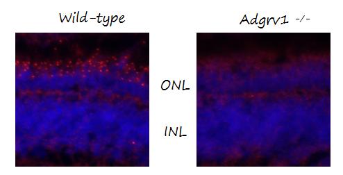
IF analysis of Ush2c/adgrv1 N-terminus using anti-Ush2c/adgrv1 N-terminus antibody .Ush2c/adgrv1 N-terminus was detected in a paraffin-embedded section of IF analysis of Ush2c/Adgrv1 using anti-Ush2c/Adgrv1 antibody .Ush2c/Adgrv1 was detected in a paraffin-embedded section of wildtype and KO mutant zebrafish retina tissue. The tissue section was blocked with 10% normal goat serum and 2% bovine serum albumin in PBS. The tissue section was then incubated with rabbit anti-Ush2c/Adgrv1 Antibody at 1:100 overnight at 4°C. Alexa Fluor 568 Conjugated Goat Anti-Rabbit IgG was used as secondary antibody at 1:800 dilution and incubated for 30 minutes at 37°C. The section was counterstained with DAPI. Visualize using a fluorescence microscope and filter sets appropriate for the label used. tissue. The tissue section was incubated with rabbit anti-Ush2c/adgrv1 N-terminus Antibody at a dilution of 1:100. Alexa Fluor 568 Conjugated Goat Anti-Rabbit IgG was used as secondary antibody. The section was counterstained with DAPI .| Product Name | Anti-Zebrafish Ush2c/adgrv1 N-terminus Antibody |
|---|---|
| Antibody Type | Primary Antibodies |
| Clonality | Polyclonal |
|---|---|
| Isotype | IgG |
| Host Species | Rabbit |
| Tested Applications | IF |
| IF:1:50-1:400 |
|
| Species Reactivity | Zebrafish |
| Concentration | 500ug/ml |
| Purification | Affinity purified |
| Target | adgrv1 |
|---|
| SwissProt ID | Q6JAN0 |
|---|
IF
IF analysis of Ush2c/adgrv1 N-terminus using anti-Ush2c/adgrv1 N-terminus antibody .Ush2c/adgrv1 N-terminus was detected in a paraffin-embedded section of IF analysis of Ush2c/Adgrv1 using anti-Ush2c/Adgrv1 antibody .Ush2c/Adgrv1 was detected in a paraffin-embedded section of wildtype and KO mutant zebrafish retina tissue. The tissue section was blocked with 10% normal goat serum and 2% bovine serum albumin in PBS. The tissue section was then incubated with rabbit anti-Ush2c/Adgrv1 Antibody at 1:100 overnight at 4°C. Alexa Fluor 568 Conjugated Goat Anti-Rabbit IgG was used as secondary antibody at 1:800 dilution and incubated for 30 minutes at 37°C. The section was counterstained with DAPI. Visualize using a fluorescence microscope and filter sets appropriate for the label used. tissue. The tissue section was incubated with rabbit anti-Ush2c/adgrv1 N-terminus Antibody at a dilution of 1:100. Alexa Fluor 568 Conjugated Goat Anti-Rabbit IgG was used as secondary antibody. The section was counterstained with DAPI .| Application Notes | IF:1:50-1:400 |
|---|
| Form | Liquid |
|---|---|
| Storage Instructions | 12 months from date of receipt,-20℃ as supplied. |
| Storage Buffer | PBS, 0.02% NaN3, 1 mg/ml BSA and 50% glycerol. |


