WB
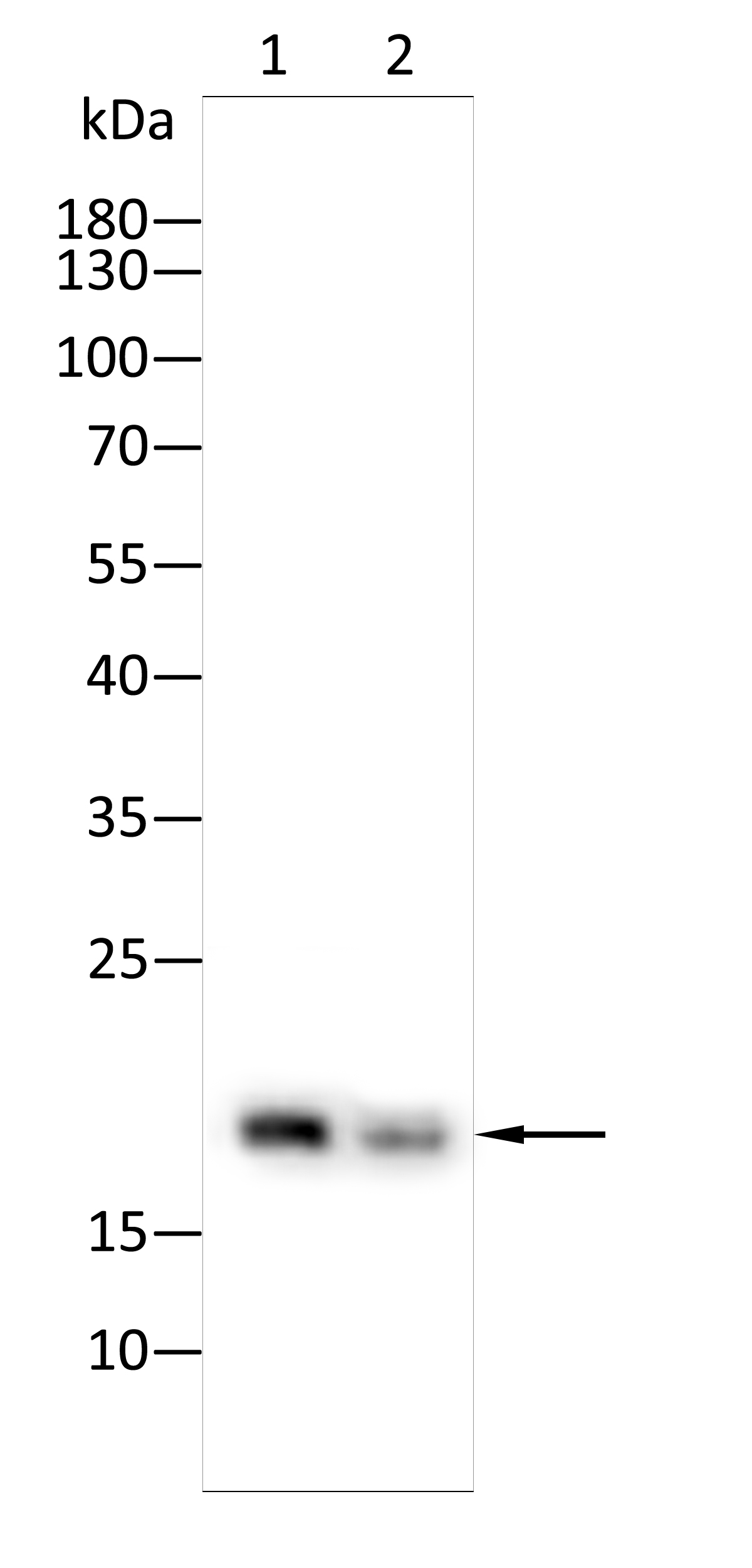
Western blot analysis using LC3A antibody against Hela(1),C2C12(2) lysate . 12% SDS-PAGE gel.Sample loading: 20μg /lane. Transfer the proteins onto a PVDF membrane (OM790003), and block it with TBST (OM750016) plus skimmed milk powder(OM750084) for one hour. Dilute the primary antibody with the antibody diluent (OM750012) at a ratio of 1:1000, and incubate it overnight at 4°C. Wash the membrane three times with TBST (OM750016), 5 minutes each time. At room temperature, dilute the secondary antibody, Goat Anti-Mouse IgG (H&L) - HRP(OM644366), at a ratio of 1:20000 and incubate for one hour. Wash the membrane three times with TBST (OM750016) again, 5 minutes each time. Use ECL (OM625701) for luminescence.staining time: 60SELISA
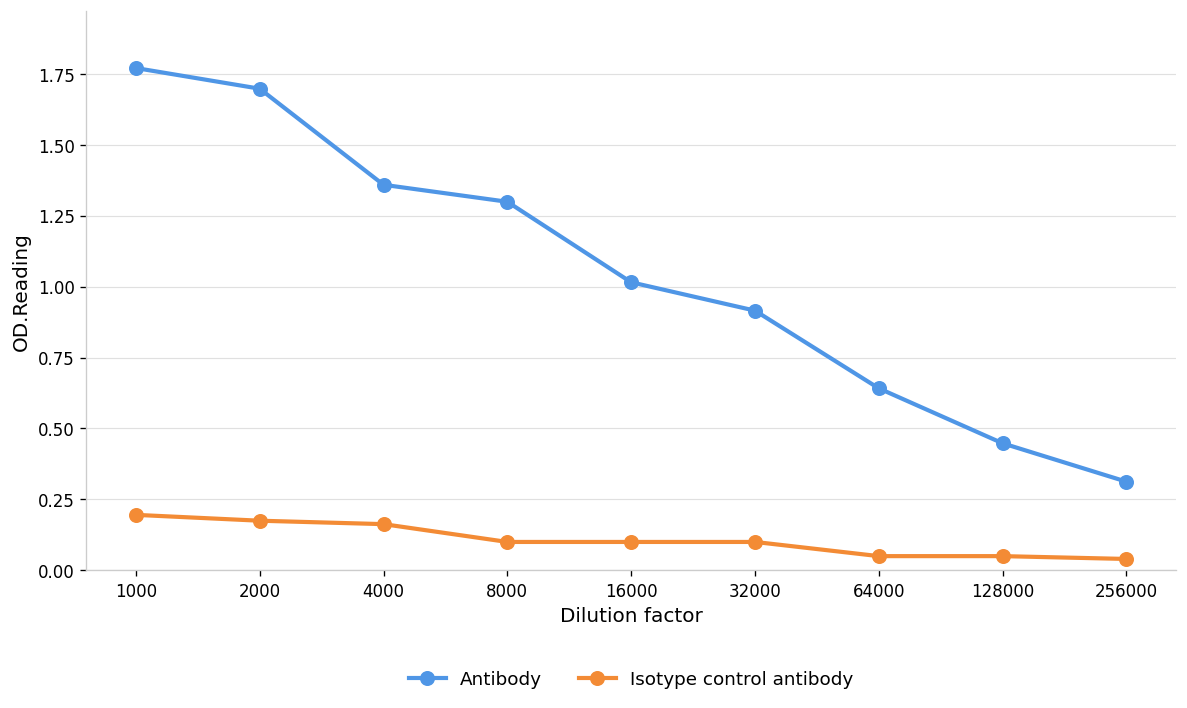
Blue line: Antibody; Orange line: Isotype control antibody| Product Name | Anti-LC3A antibody,pbs only |
|---|---|
| Antibody Type | Primary Antibodies |
| Immunogen | Recombinant protein |
| Clonality | Monoclonal |
|---|---|
| Isotype | IgG |
| Host Species | Mouse |
| Tested Applications | ELISAWB |
| WB:1:1000-1:2000 |
|
| Species Reactivity | HumanMouseRat |
| Concentration | 1mg/ml |
| Purification | Affinity purified |
| Gene Symbol | MAP1LC3A |
|---|---|
| Gene Synonyms | 1010001H21Rik 4922501H04Rik ATG8E LC3 LC3-I LC3-II LC3A MAP1ALC3 MAP1BLC3 zgc:77094 |
| Gene Full Name | microtubule associated protein 1 light chain 3 alpha |
| Gene Summary | MAP1A and MAP1B are microtubule-associated proteins which mediate the physical interactions between microtubules and components of the cytoskeleton. MAP1A and MAP1B each consist of a heavy chain subunit and multiple light chain subunits. The protein encoded by this gene is one of the light chain subunits and can associate with either MAP1A or MAP1B. Two transcript variants encoding different isoforms have been found for this gene. The expression of variant 1 is suppressed in many tumor cell lines, suggesting that may be involved in carcinogenesis. [provided by RefSeq, Feb 2012] |
| Alternative Names | autophagy-related protein LC3 A autophagy-related ubiquitin-like modifier LC3 A LC3-like protein 1 lc3a MAP1 light chain 3-like protein 1 MAP1A/1B light chain 3 A MAP1A/MAP1B LC3 A MAP1A/MAP1B light chain 3 A Microtubule-asso microtubule-associated protein 1-light chain 3A microtubule-associated proteins 1A/1B light chain 3 microtubule-associated proteins 1A/1B light chain 3A MLP3A RP11-346K17.1 |
| Molecular Weight(MW) | 15(observed:18kDa) |
| Cellular Localization | Cytoplasmic vesicle, autophagosome membrane ; Lipid-anchor . Endomembrane system ; Lipid-anchor . Cytoplasm,cytoskeleton .LC3-II binds to the autophagic membranes. |
| SwissProt ID | Q9H492 |
|---|
WB
Western blot analysis using LC3A antibody against Hela(1),C2C12(2) lysate . 12% SDS-PAGE gel.Sample loading: 20μg /lane. Transfer the proteins onto a PVDF membrane (OM790003), and block it with TBST (OM750016) plus skimmed milk powder(OM750084) for one hour. Dilute the primary antibody with the antibody diluent (OM750012) at a ratio of 1:1000, and incubate it overnight at 4°C. Wash the membrane three times with TBST (OM750016), 5 minutes each time. At room temperature, dilute the secondary antibody, Goat Anti-Mouse IgG (H&L) - HRP(OM644366), at a ratio of 1:20000 and incubate for one hour. Wash the membrane three times with TBST (OM750016) again, 5 minutes each time. Use ECL (OM625701) for luminescence.staining time: 60S
ELISA
Blue line: Antibody; Orange line: Isotype control antibody| Application Notes | WB:1:1000-1:2000 |
|---|
| Form | Liquid |
|---|---|
| Storage Instructions | Shipped at 4°C. Store at +4°C short term (1-2 weeks). Store at -20°C long term. Avoid freeze / thaw cycle. |
| Storage Buffer | PBS ONLY |


