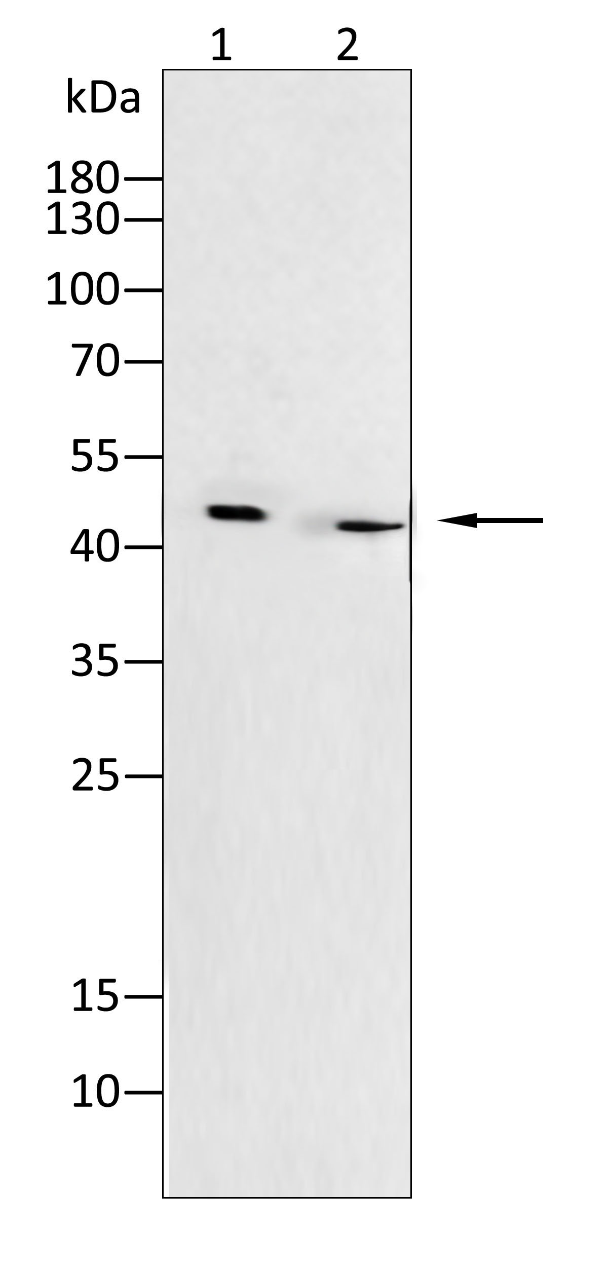
WB
Western blot analysis using Cytokeratin1 antibody against Hela(1), A431(2) cell lysate.IHC
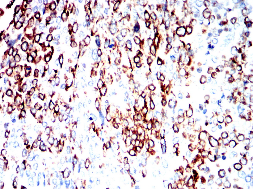
Immunohistochemical analysis of paraffin-embedded ovarian cancer tissues using Cytokeratin17 antibody with DAB staining.ICC/IF
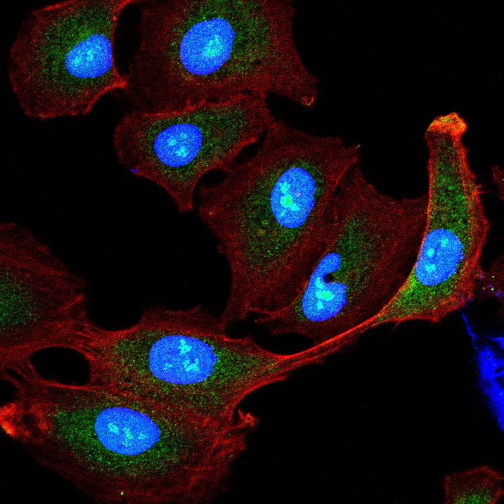
Immunofluorescence analysis of A549 cells using Cytokeratin17 antibody (green). Blue: DAPI fluorescent DNA dye. Red: Actin filaments have been labeled with Alexa Fluor- 555 phalloidin.| Product Name | Anti-Cytokeratin17 antibody |
|---|---|
| Antibody Type | Primary Antibodies |
| Immunogen | Polypeptide |
| Clonality | Polyclonal |
|---|---|
| Isotype | IgG |
| Host Species | Rabbit |
| Tested Applications | ELISAICC/IFIHCWB |
| WB:1:200-1:2000 IHC:1:200-1:1000 ICC/IF:1:100-1:500 |
|
| Species Reactivity | HumanMouseRat |
| Concentration | 1mg/ml |
| Purification | Protein A |
| Gene Symbol | KRT17 |
|---|---|
| Gene Synonyms | PC K17 PC2 39.1 CK-17 PCHC1 |
| Gene Full Name | keratin 17 |
| Gene Summary | This gene encodes the type I intermediate filament chain keratin 17, expressed in nail bed, hair follicle, sebaceous glands, and other epidermal appendages. Mutations in this gene lead to Jackson-Lawler type pachyonychia congenita and steatocystoma multiplex. [provided by RefSeq, Aug 2008] |
| Molecular Weight(MW) | 48 kDa |
| Source | Rabbit |
| Cellular Localization | Cytoplasm |
WB
Western blot analysis using Cytokeratin1 antibody against Hela(1), A431(2) cell lysate.
IHC
Immunohistochemical analysis of paraffin-embedded ovarian cancer tissues using Cytokeratin17 antibody with DAB staining.
ICC/IF
Immunofluorescence analysis of A549 cells using Cytokeratin17 antibody (green). Blue: DAPI fluorescent DNA dye. Red: Actin filaments have been labeled with Alexa Fluor- 555 phalloidin.| Application Notes | WB:1:200-1:2000 IHC:1:200-1:1000 ICC/IF:1:100-1:500 |
|---|
| Form | Liquid |
|---|---|
| Storage Instructions | Shipped at 4°C. Store at +4°C short term (1-2 weeks). Store at -20°C long term. Avoid freeze / thaw cycle. |
| Storage Buffer | Purified antibody in PBS with 0.05% sodium azide. |


 Data sheet for OM642119