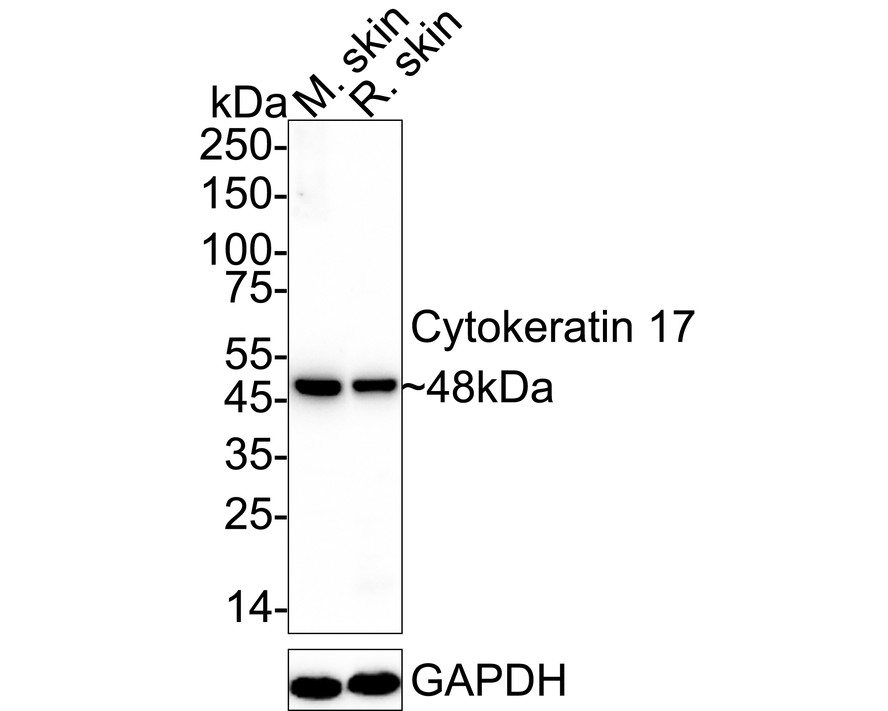
WB
Western blot analysis of Cytokeratin 17 on different lysates with Rabbit anti-Cytokeratin 17 antibody at 1/1,000 dilution. Lane 1: Mouse skin tissue lysate Lane 2: Rat skin tissue lysate Lysates/proteins at 40 µg/Lane. Exposure time: 3 minutes; 4-20% SDS-PAGE gel. Proteins were transferred to a PVDF membrane and blocked with 5% NFDM/TBST for 1 hour at room temperature. The primary antibody at 1/1,000 dilution was used in 5% NFDM/TBST at 4℃ overnight. Goat Anti-Rabbit IgG - HRP Secondary Antibody at 1/50,000 dilution was used for 1 hour at room temperature.IHC
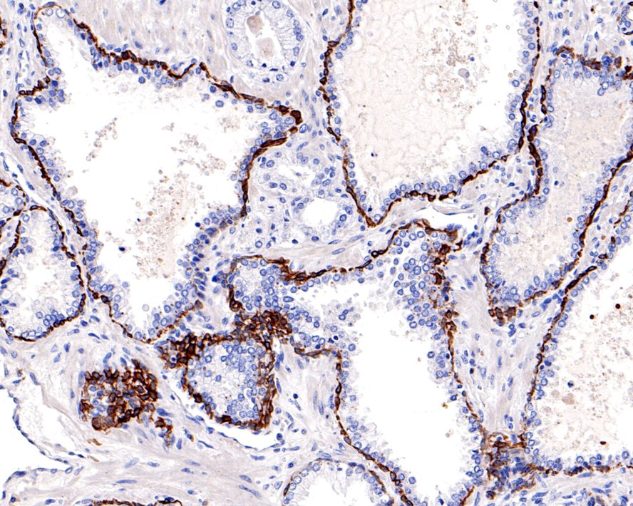
Immunohistochemical analysis of paraffin-embedded human prostate tissue using anti-Cytokeratin 17 antibody. The section was pre-treated using heat mediated antigen retrieval with Tris-EDTA buffer (pH 9.0) for 20 minutes.The tissues were blocked in 5% BSA for 30 minutes at room temperature, washed with ddH2O and PBS, and then probed with the primary antibody (ET1602-16, 1/400) for 30 minutes at room temperature. The detection was performed using an HRP conjugated compact polymer system. DAB was used as the chromogen. Tissues were counterstained with hematoxylin and mounted with DPX.ICC/IF
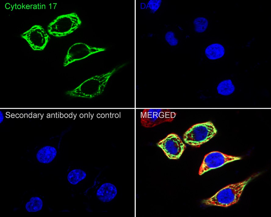
Immunocytochemistry analysis of SiHa cells labeling Cytokeratin 17 with Rabbit anti-Cytokeratin 17 antibody at 1/1,000 dilution. Cells were fixed in 4% paraformaldehyde for 15 minutes at room temperature, permeabilized with 0.1% Triton X-100 in PBS for 15 minutes at room temperature, then blocked with 1% BSA in 10% negative goat serum for 1 hour at room temperature. Cells were then incubated with Rabbit anti-Cytokeratin 17 antibody at 1/1,000 dilution in 1% BSA in PBST overnight at 4 ℃. Goat Anti-Rabbit IgG H&L (iFluor™ 488) was used as the secondary antibody at 1/1,000 dilution. PBS instead of the primary antibody was used as the secondary antibody only control. Nuclear DNA was labelled in blue with DAPI. Beta tubulin (red) was stained at 1/100 dilution overnight at +4℃. Goat Anti-Mouse IgG H&L (iFluor™ 594) was used as the secondary antibody at 1/1,000 dilution.IF-P
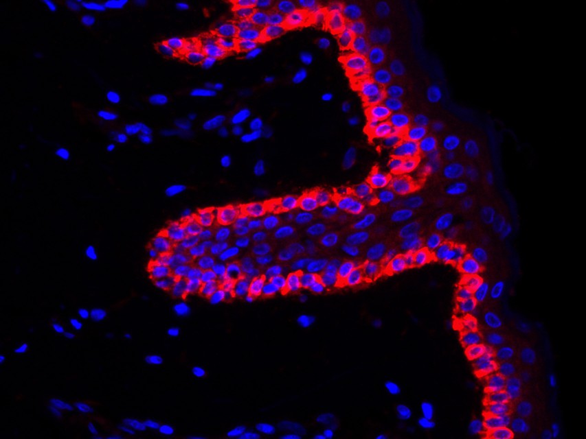
Immunofluorescence analysis of paraffin-embedded human skin tissue labeling Cytokeratin 17. The section was pre-treated using heat mediated antigen retrieval with Tris-EDTA buffer (pH 9.0) for 20 minutes. The tissues were blocked in 10% negative goat serum for 1 hour at room temperature, washed with PBS. And then probed with the primary antibodies Cytokeratin 17 (red) at 1/200 dilution at +4℃ overnight, washed with PBS. Goat Anti-Rabbit IgG H&L (iFluor™ 594) was used as the secondary antibodies at 1/1,000 dilution. Nuclei were counterstained with DAPI (blue).| Product Name | Cytokeratin 17 Recombinant Rabbit Monoclonal Antibody |
|---|---|
| Antibody Type | Primary Antibodies |
| Immunogen | Synthetic peptide within Human Cytokeratin 17 aa 1-50 / 432. |
| Clonality | Monoclonal |
|---|---|
| Isotype | IgG |
| Host Species | Rabbit |
| Tested Applications | ICC/IFIF-PIHCWB |
| WB:1:1000-1:2000 IHC:1:50-1:1500 ICC:1:50 IF-P:1:200 |
|
| Species Reactivity | HumanMouseRat |
| Concentration | 1mg/ml |
| Purification | Protein A |
| Gene Symbol | KRT17 |
|---|---|
| Gene Synonyms | PC K17 PC2 39.1 CK-17 PCHC1 |
| Gene Full Name | keratin 17 |
| Gene Summary | This gene encodes the type I intermediate filament chain keratin 17, expressed in nail bed, hair follicle, sebaceous glands, and other epidermal appendages. Mutations in this gene lead to Jackson-Lawler type pachyonychia congenita and steatocystoma multiplex. [provided by RefSeq, Aug 2008] |
| Molecular Weight(MW) | 48kDa |
| Cellular Localization | Cytoplasm. |
WB
Western blot analysis of Cytokeratin 17 on different lysates with Rabbit anti-Cytokeratin 17 antibody at 1/1,000 dilution. Lane 1: Mouse skin tissue lysate Lane 2: Rat skin tissue lysate Lysates/proteins at 40 µg/Lane. Exposure time: 3 minutes; 4-20% SDS-PAGE gel. Proteins were transferred to a PVDF membrane and blocked with 5% NFDM/TBST for 1 hour at room temperature. The primary antibody at 1/1,000 dilution was used in 5% NFDM/TBST at 4℃ overnight. Goat Anti-Rabbit IgG - HRP Secondary Antibody at 1/50,000 dilution was used for 1 hour at room temperature.
IHC
Immunohistochemical analysis of paraffin-embedded human prostate tissue using anti-Cytokeratin 17 antibody. The section was pre-treated using heat mediated antigen retrieval with Tris-EDTA buffer (pH 9.0) for 20 minutes.The tissues were blocked in 5% BSA for 30 minutes at room temperature, washed with ddH2O and PBS, and then probed with the primary antibody (ET1602-16, 1/400) for 30 minutes at room temperature. The detection was performed using an HRP conjugated compact polymer system. DAB was used as the chromogen. Tissues were counterstained with hematoxylin and mounted with DPX.
ICC/IF
Immunocytochemistry analysis of SiHa cells labeling Cytokeratin 17 with Rabbit anti-Cytokeratin 17 antibody at 1/1,000 dilution. Cells were fixed in 4% paraformaldehyde for 15 minutes at room temperature, permeabilized with 0.1% Triton X-100 in PBS for 15 minutes at room temperature, then blocked with 1% BSA in 10% negative goat serum for 1 hour at room temperature. Cells were then incubated with Rabbit anti-Cytokeratin 17 antibody at 1/1,000 dilution in 1% BSA in PBST overnight at 4 ℃. Goat Anti-Rabbit IgG H&L (iFluor™ 488) was used as the secondary antibody at 1/1,000 dilution. PBS instead of the primary antibody was used as the secondary antibody only control. Nuclear DNA was labelled in blue with DAPI. Beta tubulin (red) was stained at 1/100 dilution overnight at +4℃. Goat Anti-Mouse IgG H&L (iFluor™ 594) was used as the secondary antibody at 1/1,000 dilution.
IF-P
Immunofluorescence analysis of paraffin-embedded human skin tissue labeling Cytokeratin 17. The section was pre-treated using heat mediated antigen retrieval with Tris-EDTA buffer (pH 9.0) for 20 minutes. The tissues were blocked in 10% negative goat serum for 1 hour at room temperature, washed with PBS. And then probed with the primary antibodies Cytokeratin 17 (red) at 1/200 dilution at +4℃ overnight, washed with PBS. Goat Anti-Rabbit IgG H&L (iFluor™ 594) was used as the secondary antibodies at 1/1,000 dilution. Nuclei were counterstained with DAPI (blue).| Application Notes | WB:1:1000-1:2000 IHC:1:50-1:1500 ICC:1:50 IF-P:1:200 |
|---|
| Form | Liquid |
|---|---|
| Storage Instructions | Store at +4℃ after thawing. Aliquot store at -20℃ or -80℃. Avoid repeated freeze / thaw cycles. |
| Storage Buffer | 1*TBS (pH7.4), 0.05% BSA, 40% Glycerol. Preservative: 0.05% Sodium Azide. |


 Data sheet for OM643210