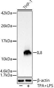
WB
Western blot analysis of lysates from THP-1 cells using IL8 Rabbit mAb at 1:1000 dilution. THP-1 cells were treated by PMA/TPA (80 nM) at 37℃ for overnight and LPS (1μg/ml) at 37℃ for 6 hours. Secondary antibody: HRP-conjugated Goat anti-Rabbit IgG (H+L) at 1:10000 dilution. Lysates/proteins: 25μg per lane. Blocking buffer: 3% nonfat dry milk in TBST. Detection: ECL Basic Kit. Exposure time: 1s.ICC/IF
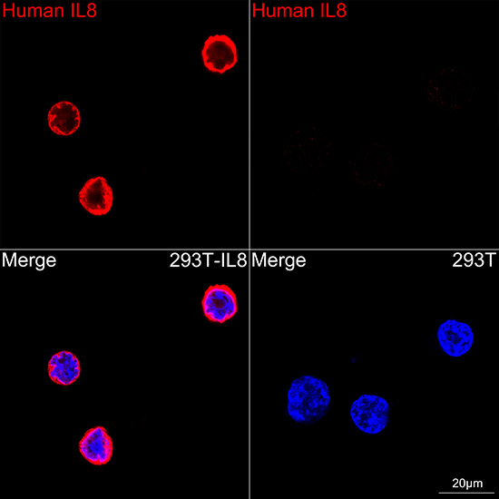
Confocal imaging of 293T cells transfected with IL8 using IL8 Rabbit mAb (dilution 1:200) followed by a further incubation with Cy3 Goat Anti-Rabbit IgG (H+L) (dilution 1:500) (Red). DAPI was used for nuclear staining (blue). Objective: 100x.FC
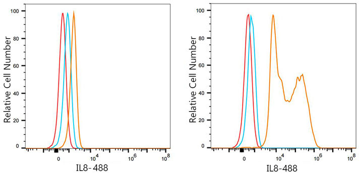
Flow cytometry: 1X10^6 293T cells (negative control,left) and 293T (Transfection,right) cells were intracellularly stained with IL8 Rabbit mAb (2ug/mL,orange line) or 488 Rabbit IgG isotype control(5 μl/Test,blue line), followed by FITC conjugated goat anti-rabbit pAb staining. Non-fluorescently stained cells were used as blank control (red line).| Product Name | IL8 Rabbit mAb |
|---|---|
| Antibody Type | Primary Antibodies |
| Immunogen | Recombinant fusion protein containing a sequence corresponding to amino acids 28-99 of human IL8(NP_000575.1). |
| Clonality | monoclonal |
|---|---|
| Isotype | IgG |
| Host Species | Rabbit |
| Tested Applications | FCICC/IFWB |
| WB:1:500-1:2000 ICC/IF:1:200-1:400 FC:1:500-1:1000 |
|
| Species Reactivity | Human |
| Concentration | 1mg/ml |
| Purification | Affinity purified |
| Gene Symbol | CXCL8 |
|---|---|
| Gene Synonyms | IL8 NAF GCP1 LECT LUCT NAP1 GCP-1 LYNAP MDNCF MONAP NAP-1 SCYB8 |
| Gene Full Name | C-X-C motif chemokine ligand 8 |
| Gene Summary | The protein encoded by this gene is a member of the CXC chemokine family and is a major mediator of the inflammatory response. The encoded protein is commonly referred to as interleukin-8 (IL-8). IL-8 is secreted by mononuclear macrophages, neutrophils, eosinophils, T lymphocytes, epithelial cells, and fibroblasts. It functions as a chemotactic factor by guiding the neutrophils to the site of infection. Bacterial and viral products rapidly induce IL-8 expression. IL-8 also participates with other cytokines in the proinflammatory signaling cascade and plays a role in systemic inflammatory response syndrome (SIRS). This gene is believed to play a role in the pathogenesis of the lower respiratory tract infection bronchiolitis, a common respiratory tract disease caused by the respiratory syncytial virus (RSV). The overproduction of this proinflammatory protein is thought to cause the lung inflammation associated with csytic fibrosis. This proinflammatory protein is also suspected of playing a role in coronary artery disease and endothelial dysfunction. This protein is also secreted by tumor cells and promotes tumor migration, invasion, angiogenesis and metastasis. This chemokine is also a potent angiogenic factor. The binding of IL-8 to one of its receptors (IL-8RB/CXCR2) increases the permeability of blood vessels and increasing levels of IL-8 are positively correlated with increased severity of multiple disease outcomes (eg, sepsis). This gene and other members of the CXC chemokine gene family form a gene cluster in a region of chromosome 4q. [provided by RefSeq, May 2020] |
| Molecular Weight(MW) | 11kDa |
| Cellular Localization | Secreted. |
WB
Western blot analysis of lysates from THP-1 cells using IL8 Rabbit mAb at 1:1000 dilution. THP-1 cells were treated by PMA/TPA (80 nM) at 37℃ for overnight and LPS (1μg/ml) at 37℃ for 6 hours. Secondary antibody: HRP-conjugated Goat anti-Rabbit IgG (H+L) at 1:10000 dilution. Lysates/proteins: 25μg per lane. Blocking buffer: 3% nonfat dry milk in TBST. Detection: ECL Basic Kit. Exposure time: 1s.
ICC/IF
Confocal imaging of 293T cells transfected with IL8 using IL8 Rabbit mAb (dilution 1:200) followed by a further incubation with Cy3 Goat Anti-Rabbit IgG (H+L) (dilution 1:500) (Red). DAPI was used for nuclear staining (blue). Objective: 100x.
FC
Flow cytometry: 1X10^6 293T cells (negative control,left) and 293T (Transfection,right) cells were intracellularly stained with IL8 Rabbit mAb (2ug/mL,orange line) or 488 Rabbit IgG isotype control(5 μl/Test,blue line), followed by FITC conjugated goat anti-rabbit pAb staining. Non-fluorescently stained cells were used as blank control (red line).| Application Notes | WB:1:500-1:2000 ICC/IF:1:200-1:400 FC:1:500-1:1000 |
|---|
| Form | Liquid |
|---|---|
| Storage Instructions | Store at -20℃. Avoid freeze / thaw cycles. |
| Storage Buffer | Buffer: PBS with 0.05% proclin300, 0.05% BSA, 50% glycerol, pH7.3. |


 Data sheet for OM644084