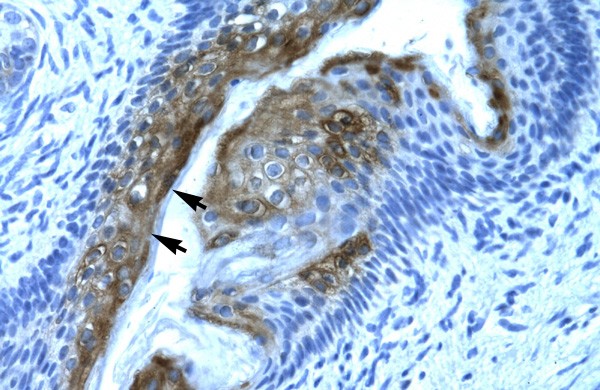


Immunohistochemical
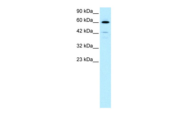
Western blot
0.2-1 ug/mlWB
The expression of HDAC2, IL-8 and VEGF proteins in tracheal tissues from rabbits after tracheal brushing and nintedanib treatments 2 day after treatments. Left panel: representative Western blots, right panel: relative protein level. * and # denote P < 0.01 vs control and model, respectively.WB
HDAC2, IL-8, TGF-β1 and VEGF expression in the tracheal tissue of each group determined by WB. Results were normalized to the internal control GAPDH. Data are presented as the mean ± standard deviation. *P<0.05 vs. CON. #P<0.05 vs. ERY. HDAC2, histone deacetylase 2; IL-8, interleukin 8; TGF-β1, transforming growth factor β1; VEGF, vascular endothelial growth factor; WB, western blotting; CON, untreated rabbit tracheal stenosis model; PEN, rabbit tracheal stenosis model treated with penicillin; ERY, rabbit tracheal stenosis model treated with erythromycin; BUD, rabbit tracheal stenosis model treated with budesonide; PEN + ERY + BUD, rabbit trachealIF
The expression of HDAC2 in the different groups determined by immunofluorescence. Image scale bar, 100 µm. Red fluorescence (Cy3 staining) for the detection of the target protein HDAC2, blue fluorescence (DAPI staining) for the nucleus. Data are presented as the mean ± standard deviation in the corresponding histogram. ∗P < 0.05 vs. the control group. Control: rabbit tracheal stenosis model without treatment; NS: rabbit tracheal stenosis model treated with penicillin; ERY: rabbit tracheal stenosis model treated with erythromycin; Budesonide: rabbit tracheal stenosis model treated with budesonide; Vorinostat: rabbit tracheal stenosis model treated with vorinostat.| Product Name | HDAC2 antibody - C-terminal region |
|---|---|
| Antibody Type | Primary Antibodies |
| Immunogen | The immunogen for anti-HDAC2 antibody: synthetic peptide directed towards the C terminal of human HDAC2 |
| Clonality | Polyclonal |
|---|---|
| Isotype | IgG |
| Host Species | Rabbit |
| Concentration | 1mg/ml |
| Purification | Affinity purified |
| Gene Symbol | HDAC2 |
|---|---|
| Gene Synonyms | HD2 RPD3 YAF1 |
| Gene Full Name | Histone deacetylase 2 |
| Gene Summary | Histone deacetylase 2 (HDAC2), or transcriptional regulator homolog RPD3 L1, is highly homologous to the yeast transcription factor RPD3 (reduced potassium dependency 3) gene. As in yeast, human HDA2 is likely to be involved in regulating chromatin structure during transcription. It has been implicated to associate with YY1, a mammalian zinc-finger transcription factor, which negatively regulates transcription by tethering RPD3 to DNA as a cofactor. This process is highly concerved from yeast to human. |
| Alternative Names | HD2 RPD3 YAF1 |
| Molecular Weight(MW) | 55kDa |
| Sequence | 488 amino acids |
Immunohistochemical
Western blot
0.2-1 ug/ml
WB
The expression of HDAC2, IL-8 and VEGF proteins in tracheal tissues from rabbits after tracheal brushing and nintedanib treatments 2 day after treatments. Left panel: representative Western blots, right panel: relative protein level. * and # denote P < 0.01 vs control and model, respectively.
WB
HDAC2, IL-8, TGF-β1 and VEGF expression in the tracheal tissue of each group determined by WB. Results were normalized to the internal control GAPDH. Data are presented as the mean ± standard deviation. *P<0.05 vs. CON. #P<0.05 vs. ERY. HDAC2, histone deacetylase 2; IL-8, interleukin 8; TGF-β1, transforming growth factor β1; VEGF, vascular endothelial growth factor; WB, western blotting; CON, untreated rabbit tracheal stenosis model; PEN, rabbit tracheal stenosis model treated with penicillin; ERY, rabbit tracheal stenosis model treated with erythromycin; BUD, rabbit tracheal stenosis model treated with budesonide; PEN + ERY + BUD, rabbit tracheal
IF
The expression of HDAC2 in the different groups determined by immunofluorescence. Image scale bar, 100 µm. Red fluorescence (Cy3 staining) for the detection of the target protein HDAC2, blue fluorescence (DAPI staining) for the nucleus. Data are presented as the mean ± standard deviation in the corresponding histogram. ∗P < 0.05 vs. the control group. Control: rabbit tracheal stenosis model without treatment; NS: rabbit tracheal stenosis model treated with penicillin; ERY: rabbit tracheal stenosis model treated with erythromycin; Budesonide: rabbit tracheal stenosis model treated with budesonide; Vorinostat: rabbit tracheal stenosis model treated with vorinostat.| Form | Liquid |
|---|---|
| Storage Instructions | Aliquot and store at -20°C. Avoid repeated freeze / thaw cycles |
| Storage Buffer | phosphate buffered saline , pH 7.4, 150mM NaCl, 0.02% sodium azide and 50% glycerol. |


