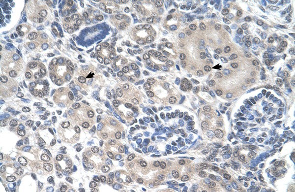
Immunohistochemical
ImmunohistochemicalWestern blot
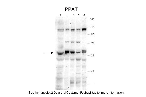
Sample Type: 1. Human Skin Fibroblasts (100ug)2. HepG2 cells (20ug)3. HEK273 cells (20ug)4. HeLa skin cells(20ug)5. Hamster CHO K1 cells (20ug)Primary Dilution: 1:5000Secondary Antibody: Clean-Blot IP detection Reagent and KitSecondary Dilution: 1:2000Image Submitted By: V. Skopova, M. ZikanovaInstitute of Inherited Metabolic Disorders, Charles University in Prague and General Hopspital in PragueWestern blot
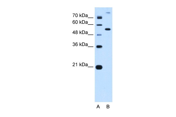
PPAT antibody - N-terminal region validated by WB using HepG2 Cell Lysate at 0.25ug/ml.| Product Name | Rabbit anti-PPAT polyclonal antibody - N-terminal region |
|---|---|
| Antibody Type | Primary Antibodies |
| Antigen Alias | ATASE, GPAT, PRAT |
| Immunogen | The immunogen for anti-PPAT antibody: synthetic peptide directed towards the N terminal of human PPAT |
| Clonality | Polyclonal |
|---|---|
| Isotype | IgG |
| Host Species | Rabbit |
| Tested Applications | WBIHC |
| WB:1:500~1:2000 IHC:1:50~1:200 Notes:Optimal dilutions/concentrations should be determined by the researcher. |
|
| Species Reactivity | BovineDogHorseHumanRabbit |
| Concentration | 1mg/ml |
| Purification | Affinity purified |
| Gene Symbol | PPAT |
|---|---|
| Gene Synonyms | ATASE GPAT PRAT |
| Gene Full Name | Phosphoribosyl pyrophosphate amidotransferase |
| Gene Summary | PPAT is a member of the purine/pyrimidine phosphoribosyltransferase family. This protein is a regulatory allosteric enzyme that catalyzes the first step of de novo purine nucleotide biosynthesis. Its gene and PAICS/AIRC, a bifunctional enzyme catalyzing steps six and seven in the purine nucleotide biosynthesis pathway, are located in close proximity on chromosome 4.The protein encoded by this gene is a member of the purine/pyrimidine phosphoribosyltransferase family. This protein is a regulatory allosteric enzyme that catalyzes the first step of de novo purine nucleotide biosynthesis. This gene and PAICS/AIRC, a bifunctional enzyme catalyzing steps six and seven in the purine nucleotide biosynthesis pathway, are located in close proximity on chromosome 4. |
| Alternative Names | ATASE GPAT PRAT |
| Molecular Weight(MW) | 56kDa |
| Sequence | 517 amino acids |
Immunohistochemical
Immunohistochemical
Western blot
Sample Type: 1. Human Skin Fibroblasts (100ug)2. HepG2 cells (20ug)3. HEK273 cells (20ug)4. HeLa skin cells(20ug)5. Hamster CHO K1 cells (20ug)Primary Dilution: 1:5000Secondary Antibody: Clean-Blot IP detection Reagent and KitSecondary Dilution: 1:2000Image Submitted By: V. Skopova, M. ZikanovaInstitute of Inherited Metabolic Disorders, Charles University in Prague and General Hopspital in Prague
Western blot
PPAT antibody - N-terminal region validated by WB using HepG2 Cell Lysate at 0.25ug/ml.| Application Notes | WB:1:500~1:2000 IHC:1:50~1:200 Notes:Optimal dilutions/concentrations should be determined by the researcher. |
|---|
| Form | Liquid |
|---|---|
| Storage Instructions | Aliquot and store at -20°C. Avoid repeated freeze / thaw cycles |
| Storage Buffer | phosphate buffered saline , pH 7.4, 150mM NaCl, 0.02% sodium azide and 50% glycerol. |


