Western blot
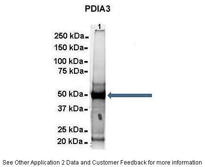
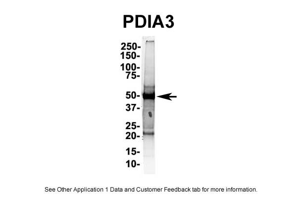
500 ug Human NT2 cell lysateAmount of IP Antibody :6 ugPrimary Antibody :PDIA3Primary Antibody Dilution :1:500Secondary Antibody :Goat anti-rabbit Alexa-Fluor 594Secondary Antibody Dilution :1:5000Gene Name :PDIA3Submitted by :Dr. Yuzhi Chen, University of Arkansas for Medical ScienceWestern blot
IP Suggested Anti-PDIA3 Antibody Positive Control: NT2 CELL/BRAIN TISSUEImmunofluorescence
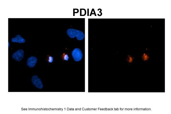
Sample Type: NT2 cellsRed: AntibodyBlue: DAPIPrimary Dilution: 1ug/50ul antibodySecondary Antibody: Alexa goat anti-rabbit 594Image Submitted by: Yuzhi Chen, University of Arkansas for Medical SciencesImmunohistochemical
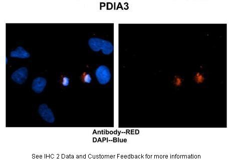
Human brain stem cellsPrimary Antibody Dilution :1:500Secondary Antibody :Goat anti-rabbit Alexa-Fluor 594Secondary Antibody Dilution :1:1000Color/Signal Descriptions :PDIA3: Red DAPI:BlueGene Name :PDIA3Submitted by :Dr. Yuzhi Chen, University of Arkansas for Medical ScienceWestern blot
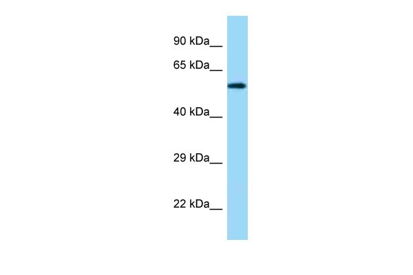
PDIA3 antibody - C-terminal region validated by WB using Fetal Kidney Lysate at 1.0ug/ml.| Product Name | Rabbit anti-PDIA3 polyclonal antibody - C-terminal region |
|---|---|
| Antibody Type | Primary Antibodies |
| Antigen Alias | ER60, ERp57, ERp60, ERp61, GRP57, GRP58, HsT17083, P58, PI-PLC |
| Clonality | Polyclonal |
|---|---|
| Isotype | IgG |
| Host Species | Rabbit |
| Tested Applications | WBIHCIP |
| WB:1:500~1:2000 IHC:1:50~1:200 IP:1:100~1:500 Notes:Optimal dilutions/concentrations should be determined by the researcher. |
|
| Species Reactivity | BovineDogGoatHumanMousePigRabbitRatSheepZebra Fish |
| Concentration | 1mg/ml |
| Purification | Affinity purified |
| Gene Symbol | PDIA3 |
|---|---|
| Gene Synonyms | ER60 ERp57 ERp60 ERp61 GRP57 GRP58 HsT17083 P58 PI-PLC |
| Gene Full Name | Protein disulfide isomerase family A, member 3 |
| Gene Summary | This gene encodes a protein of the endoplasmic reticulum that interacts with lectin chaperones calreticulin and calnexin to modulate folding of newly synthesized glycoproteins. The protein was once thought to be a phospholipase; however, it has been demonstrated that the protein actually has protein disulfide isomerase activity. It is thought that complexes of lectins and this protein mediate protein folding by promoting formation of disulfide bonds in their glycoprotein substrates. |
| Alternative Names | ER60 ERp57 ERp60 ERp61 GRP57 GRP58 HsT17083 P58 PI-PLC |
| Molecular Weight(MW) | 56kDa |
| Sequence | 505 amino acids |
Western blot
500 ug Human NT2 cell lysateAmount of IP Antibody :6 ugPrimary Antibody :PDIA3Primary Antibody Dilution :1:500Secondary Antibody :Goat anti-rabbit Alexa-Fluor 594Secondary Antibody Dilution :1:5000Gene Name :PDIA3Submitted by :Dr. Yuzhi Chen, University of Arkansas for Medical Science
Western blot
IP Suggested Anti-PDIA3 Antibody Positive Control: NT2 CELL/BRAIN TISSUE
Immunofluorescence
Sample Type: NT2 cellsRed: AntibodyBlue: DAPIPrimary Dilution: 1ug/50ul antibodySecondary Antibody: Alexa goat anti-rabbit 594Image Submitted by: Yuzhi Chen, University of Arkansas for Medical Sciences
Immunohistochemical
Human brain stem cellsPrimary Antibody Dilution :1:500Secondary Antibody :Goat anti-rabbit Alexa-Fluor 594Secondary Antibody Dilution :1:1000Color/Signal Descriptions :PDIA3: Red DAPI:BlueGene Name :PDIA3Submitted by :Dr. Yuzhi Chen, University of Arkansas for Medical Science
Western blot
PDIA3 antibody - C-terminal region validated by WB using Fetal Kidney Lysate at 1.0ug/ml.| Application Notes | WB:1:500~1:2000 IHC:1:50~1:200 IP:1:100~1:500 Notes:Optimal dilutions/concentrations should be determined by the researcher. |
|---|
| Form | Liquid |
|---|---|
| Storage Instructions | Aliquot and store at -20°C. Avoid repeated freeze / thaw cycles |
| Storage Buffer | phosphate buffered saline , pH 7.4, 150mM NaCl, 0.02% sodium azide and 50% glycerol. |


