Application
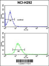
Flow cytometric analysis of NCI-H292 cells (bottom histogram) compared to a negative control cell (top histogram).FITC-conjugated goat-anti-rabbit secondary antibodies were used for the analysis.| Product Name | PDIA3 Antibody |
|---|---|
| Antibody Type | Primary Antibodies |
| Antigen Alias | Protein disulfide-isomerase A3, 58 kDa glucose-regulated protein, 58 kDa microsomal protein, p58, Disulfide isomerase ER-60, Endoplasmic reticulum resident protein 57, ER protein 57, ERp57, Endoplasmic reticulum resident protein 60, ER protein 60, ERp60, P |
| Product description | PDIA3 is the endoplasmic reticulum that interacts with lectin chaperones calreticulin and calnexin to modulate folding of newly synthesized glycoproteins. The protein was once thought to be a phospholipase; however, it has been demonstrated that the protein actually has protein disulfide isomerase activity. It is thought that complexes of lectins and this protein mediate protein folding by promoting formation of disulfide bonds in their glycoprotein substrates.1) Vigneron,N., et.al., Eur. J. Immunol. 39 (9), 2371-2376 (2009) |
| Immunogen | This PDIA3 antibody is generated from rabbits immunized with a KLH conjugated synthetic peptide between 446-475 amino acids from the C-terminal region of human PDIA3. |
| Clonality | Polyclonal |
|---|---|
| Isotype | Ig |
| Host Species | Rabbit |
| Tested Applications | FACSIHC-PWB |
| For WB starting dilution is: 1:1000 For IHC-P starting dilution is: 1:50~100 For FACS starting dilution is: 1:10~50: |
|
| Species Reactivity | Human |
| Concentration | 1mg/ml |
| Gene Symbol | PDIA3 |
|---|---|
| Alternative Names | Protein disulfide-isomerase A3 58 kDa glucose-regulated protein 58 kDa microsomal protein p58 Disulfide isomerase ER-60 Endoplasmic reticulum resident protein 57 ER protein 57 ERp57 Endoplasmic reticulum resident protein 60 ER protein 60 ERp60 P |
| Molecular Weight(MW) | 57 kDa |
Application
Flow cytometric analysis of NCI-H292 cells (bottom histogram) compared to a negative control cell (top histogram).FITC-conjugated goat-anti-rabbit secondary antibodies were used for the analysis.| Application Notes | For WB starting dilution is: 1:1000 For IHC-P starting dilution is: 1:50~100 For FACS starting dilution is: 1:10~50: |
|---|
| Form | Liquid |
|---|---|
| Storage Instructions | Store at 4˚C for three months and -20˚C, stable for up to one year. As with all antibodies care should be taken to avoid repeated freeze thaw cycles. Antibodies should not be exposed to prolonged high temperatures. |
| Storage Buffer | Supplied in PBS with 0.09% (W/V) sodium azide. |


