Immunofluorescence
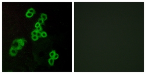
Immunofluorescence analysis of MCF7 cells, using EDG2 Antibody. The picture on the right is treated with the synthesized peptide.Western blot
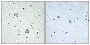
Immunohistochemistry analysis of paraffin-embedded human brain tissue, using EDG2 Antibody. The picture on the right is treated with the synthesized peptide.Western blot analysis of extracts from Jurkat cells, using EDG2 Antibody. The lane on the right is treated with the synthesized peptideWB
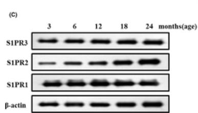
C, Western blot images and their quantification of sphingosine-1-phosphate receptor-3 (S1PR1-3) accumulation in the colon muscle layers from different groups of rats.IF
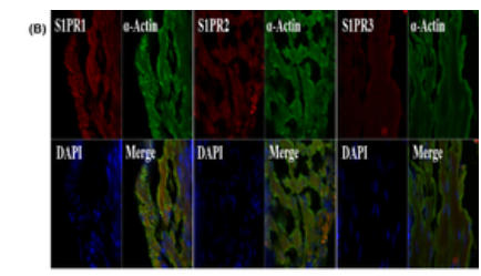
B, Representative double immunofluorescence staining of colon smooth muscle (CSM) for α-actin and sphingosine-1-phosphate receptor-3 (S1PR1-3). Nuclei were identified by DAPI staining.| Product Name | Rabbit anti-EDG2 polyclonal antibody |
|---|---|
| Antibody Type | Primary Antibodies |
| Product description |
|
| Immunogen | The antiserum was produced against synthesized peptide derived from human EDG2. |
| Clonality | Polyclonal |
|---|---|
| Host Species | Rabbit |
| Tested Applications | ELISAIFIHCWB |
| WB:1:500~1:1000 IHC:1:50~1:100 IF:1:100~1:500 ELISA:1:1000 |
|
| Species Reactivity | HumanRat |
| Concentration | 1mg/ml |
| Purification | Affinity purified |
| Gene Symbol | LPAR1 |
|---|---|
| Gene Synonyms | Lysophosphatidic acid receptor Edg-2 LPA receptor 1 LPA-1 EDG2 LPA1 |
Immunofluorescence
Immunofluorescence analysis of MCF7 cells, using EDG2 Antibody. The picture on the right is treated with the synthesized peptide.
Western blot
Immunohistochemistry analysis of paraffin-embedded human brain tissue, using EDG2 Antibody. The picture on the right is treated with the synthesized peptide.Western blot analysis of extracts from Jurkat cells, using EDG2 Antibody. The lane on the right is treated with the synthesized peptide
WB
C, Western blot images and their quantification of sphingosine-1-phosphate receptor-3 (S1PR1-3) accumulation in the colon muscle layers from different groups of rats.
IF
B, Representative double immunofluorescence staining of colon smooth muscle (CSM) for α-actin and sphingosine-1-phosphate receptor-3 (S1PR1-3). Nuclei were identified by DAPI staining.| Application Notes | WB:1:500~1:1000 IHC:1:50~1:100 IF:1:100~1:500 ELISA:1:1000 |
|---|
| Form | Liquid |
|---|---|
| Storage Instructions | store at -20°C. Avoid repeated freeze / thaw cycles |
| Storage Buffer | Rabbit IgG in phosphate buffered saline (without Mg2+ and Ca2+), pH 7.4, 150mM NaCl, 0.02% sodium azide and 50% glycerol. |


