WB
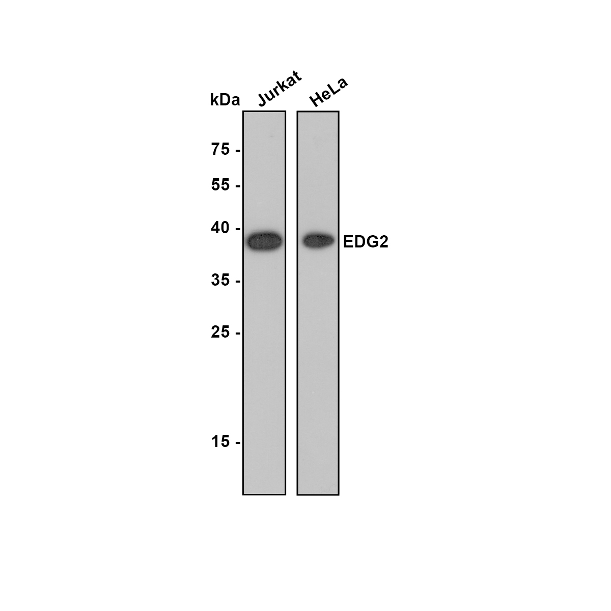
Western blot analysis of EDG2 on different lysates. Proteins were transferred to a PVDF membrane and blocked with 5% NFDM/TBST for 1 hour at room temperature. The primary antibody (1/500) was used in 5% NFDM/TBST at room temperature for 2 hours. Goat Anti-Rabbit IgG - HRP Secondary Antibody at 1/200,000 dilution was used for 1 hour at room temperature. Positive control:IHC
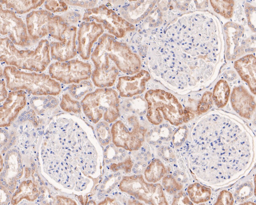
Immunohistochemical analysis of paraffin-embedded human kidney tissue using anti-EDG2 antibody. The section was pre-treated using heat mediated antigen retrieval with Tris-EDTA buffer (pH 9.0) for 20 minutes.The tissues were blocked in 5% BSA for 30 minutes at room temperature, washed with ddH2O and PBS, and then probed with the primary antibody (1/200) for 30 minutes at room temperature. The detection was performed using an HRP conjugated compact polymer system. DAB was used as the chromogen. Tissues were counterstained with hematoxylin and mounted with DPX.IHC
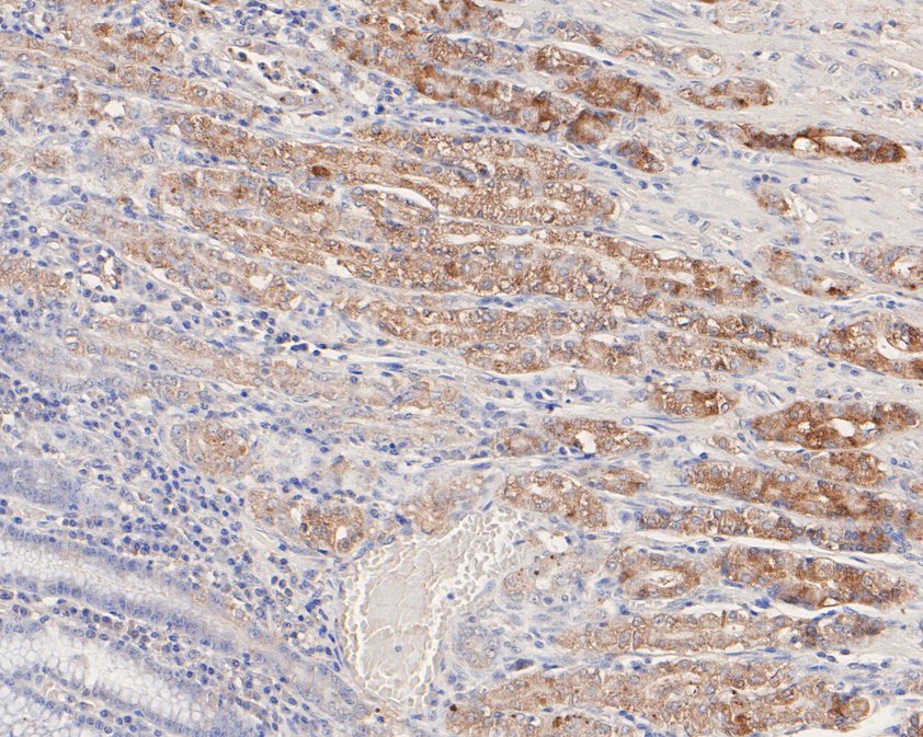
Immunohistochemical analysis of paraffin-embedded human stomach tissue using anti-EDG2 antibody. The section was pre-treated using heat mediated antigen retrieval with Tris-EDTA buffer (pH 9.0) for 20 minutes.The tissues were blocked in 5% BSA for 30 minutes at room temperature, washed with ddH2O and PBS, and then probed with the primary antibody (1/50) for 30 minutes at room temperature. The detection was performed using an HRP conjugated compact polymer system. DAB was used as the chromogen. Tissues were counterstained with hematoxylin and mounted with DPX.IHC
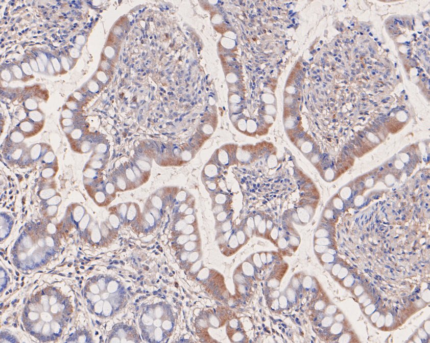
Immunohistochemical analysis of paraffin-embedded human small intestine tissue using anti-EDG2 antibody. The section was pre-treated using heat mediated antigen retrieval with Tris-EDTA buffer (pH 9.0) for 20 minutes.The tissues were blocked in 5% BSA for 30 minutes at room temperature, washed with ddH2O and PBS, and then probed with the primary antibody (1/50) for 30 minutes at room temperature. The detection was performed using an HRP conjugated compact polymer system. DAB was used as the chromogen. Tissues were counterstained with hematoxylin and mounted with DPX.FC
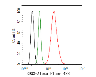
Flow cytometric analysis of EDG2 was done on Hela cells. The cells were fixed, permeabilized and stained with the primary antibody (1ug/ml) (red) compared with Rabbit IgG, monoclonal - Isotype Control (green). After incubation of the primary antibody at +4℃ for 1 hour, the cells were stained with a Alexa Fluor®488 conjugate-Goat anti-Rabbit IgG Secondary antibody at 1/1,000 dilution for 30 minutes at +4℃ (dark incubation).Unlabelled sample was used as a control (cells without incubation with primary antibody; black).| Product Name | EDG2 Recombinant Rabbit Monoclonal Antibody |
|---|---|
| Antibody Type | Primary Antibodies |
| Immunogen | Synthetic peptide within human EDG2 aa 1-50/364. |
| Clonality | Monoclonal |
|---|---|
| Isotype | IgG |
| Host Species | Rabbit |
| Tested Applications | FCIHCWB |
| WB:1:500-1:2000 IHC:1:50-1:200 FC:1:500-1:1000 |
|
| Species Reactivity | Human |
| Concentration | 1mg/ml |
| Purification | Protein A |
| Gene Symbol | LPAR1 |
|---|---|
| Gene Synonyms | EDG2 LPA1 VZG1 edg-2 vzg-1 Gpcr26 Mrec1.3 rec.1.3 |
| Gene Full Name | lysophosphatidic acid receptor 1 |
| Gene Summary | The integral membrane protein encoded by this gene is a lysophosphatidic acid (LPA) receptor from a group known as EDG receptors. These receptors are members of the G protein-coupled receptor superfamily. Utilized by LPA for cell signaling, EDG receptors mediate diverse biologic functions, including proliferation, platelet aggregation, smooth muscle contraction, inhibition of neuroblastoma cell differentiation, chemotaxis, and tumor cell invasion. Many transcript variants encoding a few different isoforms have been identified for this gene. [provided by RefSeq, Oct 2020] |
| Alternative Names | 5031439C20 antibody AI326300 antibody EDG 2 antibody EDG2 antibody Endothelial differentiation gene 2 antibody Endothelial differentiation lysophosphatidic acid G protein coupled receptor 2 antibody GPCR 26 antibody Gpcr26 antibody Gpcr91 antibody GPR26 antibody 5031439C20 antibody AI326300 antibody EDG 2 antibody EDG2 antibody Endothelial differentiation gene 2 antibody Endothelial differentiation lysophosphatidic acid G protein coupled receptor 2 antibody GPCR 26 antibody Gpcr26 antibody Gpcr91 antibody GPR26 antibody Kdt2 antibody LPA 1 antibody LPA receptor 1 antibody LPA receptor EDG2 antibody LPA-1 antibody lpA1 antibody Lpar1 antibody LPAR1_HUMAN antibody Lysophosphatidic acid receptor 1 antibody Lysophosphatidic acid receptor Edg-2 antibody Lysophosphatidic acid receptor EDG2 antibody MGC105279 antibody MGC29102 antibody Mrec1.3 antibody rec.1.3 antibody Ventricular zone gene 1 antibody vzg-1 antibody VZG1 antibody |
| Molecular Weight(MW) | 41 kDa |
| Cellular Localization | Cell membrane, Endosome, Cell surface. |
WB
Western blot analysis of EDG2 on different lysates. Proteins were transferred to a PVDF membrane and blocked with 5% NFDM/TBST for 1 hour at room temperature. The primary antibody (1/500) was used in 5% NFDM/TBST at room temperature for 2 hours. Goat Anti-Rabbit IgG - HRP Secondary Antibody at 1/200,000 dilution was used for 1 hour at room temperature. Positive control:
IHC
Immunohistochemical analysis of paraffin-embedded human kidney tissue using anti-EDG2 antibody. The section was pre-treated using heat mediated antigen retrieval with Tris-EDTA buffer (pH 9.0) for 20 minutes.The tissues were blocked in 5% BSA for 30 minutes at room temperature, washed with ddH2O and PBS, and then probed with the primary antibody (1/200) for 30 minutes at room temperature. The detection was performed using an HRP conjugated compact polymer system. DAB was used as the chromogen. Tissues were counterstained with hematoxylin and mounted with DPX.
IHC
Immunohistochemical analysis of paraffin-embedded human stomach tissue using anti-EDG2 antibody. The section was pre-treated using heat mediated antigen retrieval with Tris-EDTA buffer (pH 9.0) for 20 minutes.The tissues were blocked in 5% BSA for 30 minutes at room temperature, washed with ddH2O and PBS, and then probed with the primary antibody (1/50) for 30 minutes at room temperature. The detection was performed using an HRP conjugated compact polymer system. DAB was used as the chromogen. Tissues were counterstained with hematoxylin and mounted with DPX.
IHC
Immunohistochemical analysis of paraffin-embedded human small intestine tissue using anti-EDG2 antibody. The section was pre-treated using heat mediated antigen retrieval with Tris-EDTA buffer (pH 9.0) for 20 minutes.The tissues were blocked in 5% BSA for 30 minutes at room temperature, washed with ddH2O and PBS, and then probed with the primary antibody (1/50) for 30 minutes at room temperature. The detection was performed using an HRP conjugated compact polymer system. DAB was used as the chromogen. Tissues were counterstained with hematoxylin and mounted with DPX.
FC
Flow cytometric analysis of EDG2 was done on Hela cells. The cells were fixed, permeabilized and stained with the primary antibody (1ug/ml) (red) compared with Rabbit IgG, monoclonal - Isotype Control (green). After incubation of the primary antibody at +4℃ for 1 hour, the cells were stained with a Alexa Fluor®488 conjugate-Goat anti-Rabbit IgG Secondary antibody at 1/1,000 dilution for 30 minutes at +4℃ (dark incubation).Unlabelled sample was used as a control (cells without incubation with primary antibody; black).| Application Notes | WB:1:500-1:2000 IHC:1:50-1:200 FC:1:500-1:1000 |
|---|
| Form | Liquid |
|---|---|
| Storage Instructions | Shipped at 4℃. Store at +4℃ short term (1-2 weeks). It is recommended to aliquot into single-use upon delivery. Store at -20℃ long term. |
| Storage Buffer | TBS (pH7.4), 0.05% BSA, 40% Glycerol. Preservative: 0.05% Sodium Azide. |


