Application
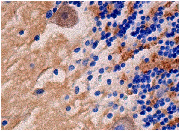
Immunoperoxidase staining of formalin fixed, paraffin-embedded human cerebellum tissue showing cytoplasmic staining of cells in granular and molecular layers and Purkinje cells.Application
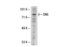
Western blot analysis of DR6 expression in Jurkat whole cell lysate.Application
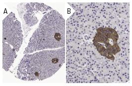
Immunoperoxidase staining of formalin fixed, paraffin-embedded human pancreas tissue showing cytoplasmic staining of islet cells at low (A) and high (B) magnification. Kindly provided by The Swedish Human Protein Atlas (HPA) program.Application
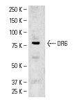
Western blot analysis of DR6 expression in K-562 whole cell lysate.| Product Name | DR6 Antibody (H-150) |
|---|---|
| Antibody Type | Primary Antibodies |
| Antigen Alias | epitope corresponding to amino acids 71-220 of DR6 of human origin |
| Modification Notes | Tumor necrosis factor (TNF) is a pleiotropic cytokine whose function is mediated by two distinct cell surface receptors, designated TNF-R1 and TNF-R2, which are expressed on most cell types. TNF function is primarily mediated through TNF-R1 signaling. Both TNF-R1 and TNF-R2 belong to the growing TNF receptor superfamily which includes the FAS antigen and CD40. TNF-R1 contains a cytoplasmic motif, termed the “death domain,” that has been found to be necessary for the transduction of the apoptotic signal. The death domain is also found in several other receptors, including FAS, DR2 (or TRUNDD), DR3 (death receptor 3), DR4, DR5 and DR6. TRUNDD, DR4 and DR5 are receptors for the apoptosis-inducing cytokine, TRAIL. A non-death domain-containing receptor, designated decoy receptor (DcRI or TRID), also specifically associates with TRAIL and may play a role in cellular resistance to apoptotic stimuli. |
| Clonality | Polyclonal |
|---|---|
| Isotype | IgG |
| Host Species | Rabbit |
| Tested Applications | WBIPIFIHCELISA |
| recommended for detection of DR6 of mouse, rat and human origin by WB, IP, IF, IHC(P) and ELISA also reactive with additional species, including equine and porcine: |
|
| Species Reactivity | HumanMouseRat |
| Concentration | 1mg/ml |
| Purification | Affinity purified |
| Alternative Names | epitope corresponding to amino acids 71-220 of DR6 of human origin |
|---|---|
| Tissue Specificity | epitope corresponding to amino acids 71-220 of DR6 of human origin |
| Entrez Gene | 27242 |
|---|
Application
Immunoperoxidase staining of formalin fixed, paraffin-embedded human cerebellum tissue showing cytoplasmic staining of cells in granular and molecular layers and Purkinje cells.
Application
Western blot analysis of DR6 expression in Jurkat whole cell lysate.
Application
Immunoperoxidase staining of formalin fixed, paraffin-embedded human pancreas tissue showing cytoplasmic staining of islet cells at low (A) and high (B) magnification. Kindly provided by The Swedish Human Protein Atlas (HPA) program.
Application
Western blot analysis of DR6 expression in K-562 whole cell lysate.| Application Notes | recommended for detection of DR6 of mouse, rat and human origin by WB, IP, IF, IHC(P) and ELISA also reactive with additional species, including equine and porcine: |
|---|
| Form | Liquid |
|---|---|
| Storage Instructions | For short-term storage, store at 4° C. For long-term storage, aliquot and store at -20ºC or below. Avoid multiple freeze-thaw cycles. |
| Storage Buffer | phosphate buffered saline , pH 7.4, 150mM NaCl, 0.02% sodium azide and 50% glycerol. |


