Application
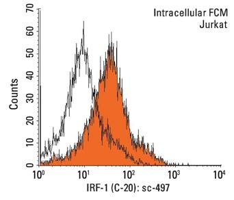
Indirect, intracellular FCM analysis of fixed and permeabilized Jurkat cells stained with IRF-1 (C-20), followed by PE-conjugated goat anti-rabbit IgG: . Black line histogram represents the isotype control, normal rabbit IgG: .Application
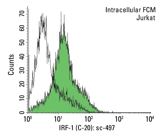
Indirect, intracellular FCM analysis of fixed and permeabilized Jurkat cells stained with IRF-1 (C-20), followed by FITC-conjugated goat anti-rabbit IgG: . Black line histogram represents the isotype control, normal rabbit IgG: .Application
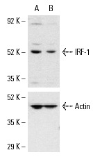
Western blot analysis of IRF-1 expression in non-transfected control (A) and IRF-1 siRNA transfected (B) HeLa cells. Blot probed with IRF-1 (C-20): . Actin (I-19): used as specificity and loading control.Application
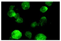
Immunofluorescence staining of methanol-fixed Jurkat cells showing nuclear staining.Application
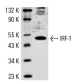
Western blot analysis of IRF-1 expression in Jurkat whole cell lysate.| Product Name | IRF-1 Antibody (C-20) |
|---|---|
| Antibody Type | Primary Antibodies |
| Antigen Alias | epitope mapping at the C-terminus of IRF-1 of human origin |
| Modification Notes | Interferon regulatory factor-1 (IRF-1) and IRF-2 have been identified as novel DNA-binding factors that function as regulators of both type I interferon (interferon-å and ∫) and interferon-inducible genes. The two factors are structurally related, particularly in their N-terminal regions, which confer DNA binding specificity. In addition, both bind to the same sequence within the promoters of interferon-å and interferon-∫ genes. IRF-1 functions as an activator of interferon transcription, while IRF-2 binds to the same cis elements and represses IRF-1 action. IRF-1 and IRF-2 have been reported to act in a mutually antagonistic manner in regulating cell growth; overexpression of the repressor IRF-2 leads to cell transformation while concomitant overexpression of IRF-1 causes reversion. IRF-1 and IRF-2 are members of a larger family of DNA binding proteins that includes IRF-3, IRF-4, IRF-5, IRF-6, IRF-7, ISGF-3© p48 (a component of the ISGF-3 complex) and IFN consensus sequence-binding protein (ICSBP). |
| Clonality | Polyclonal |
|---|---|
| Isotype | IgG |
| Host Species | Rabbit |
| Tested Applications | WBIPIFFCELISA |
| recommended for detection of IRF-1 of human origin by WB, IP, IF, FCM, ELISA also reactive with additional species, including equine, canine, bovine and porcine: |
|
| Species Reactivity | HumanMouseRat |
| Concentration | 1mg/ml |
| Purification | Affinity purified |
| Alternative Names | epitope mapping at the C-terminus of IRF-1 of human origin |
|---|---|
| Tissue Specificity | epitope mapping at the C-terminus of IRF-1 of human origin |
| Entrez Gene | 3659 |
|---|
Application
Indirect, intracellular FCM analysis of fixed and permeabilized Jurkat cells stained with IRF-1 (C-20), followed by PE-conjugated goat anti-rabbit IgG: . Black line histogram represents the isotype control, normal rabbit IgG: .
Application
Indirect, intracellular FCM analysis of fixed and permeabilized Jurkat cells stained with IRF-1 (C-20), followed by FITC-conjugated goat anti-rabbit IgG: . Black line histogram represents the isotype control, normal rabbit IgG: .
Application
Western blot analysis of IRF-1 expression in non-transfected control (A) and IRF-1 siRNA transfected (B) HeLa cells. Blot probed with IRF-1 (C-20): . Actin (I-19): used as specificity and loading control.
Application
Immunofluorescence staining of methanol-fixed Jurkat cells showing nuclear staining.
Application
Western blot analysis of IRF-1 expression in Jurkat whole cell lysate.| Application Notes | recommended for detection of IRF-1 of human origin by WB, IP, IF, FCM, ELISA also reactive with additional species, including equine, canine, bovine and porcine: |
|---|
| Form | Liquid |
|---|---|
| Storage Instructions | For short-term storage, store at 4° C. For long-term storage, aliquot and store at -20ºC or below. Avoid multiple freeze-thaw cycles. |
| Storage Buffer | phosphate buffered saline , pH 7.4, 150mM NaCl, 0.02% sodium azide and 50% glycerol. |


